A caesarean section is an incision through the abdominal wall to deliver a calf or other animal. Caesarean sections, also known as C-sections, are necessary in adult dairy cattle when vaginal delivery is too difficult for the cow or could endanger her life or the life of her calf.
Indications for Caesarean Sections
Caesarean section is potentially indicated in cases of dystocia when a calf cannot be delivered by fetal mutation and extraction. The decision on whether to perform a fetotomy or a cesarean section is dictated by whether the calf is alive, the availability of operating space in the cow’s pelvis, whether the cervix is open, access to restraint facilities, perceived value of the cow, perceived value of the calf, and the importance of future cow fertility. C-sections are commonly performed when calves are oversized (too large), which is common when the heifer (first time mother) is immature, and for embryo transfer calves. Other indications include inadequate cervical dilation (not enough relaxation of the cervix muscles), abnormal pelvic bone conformation (shape) in the cow, rupture of the cow’s abdominal musculature, problems with uterine position or uterine function, abnormalities of the cow’s uterus or vagina, abnormal calf position that is not correctable in the vagina, fetal monsters and presence of a dead fetus.
Anesthesia
General anesthesia
In sheep general anesthesia may be used, for example using Saffan (alfadalone/alfaxalone), or halothane or xylazine. This provides good relaxation and anesthesia, and avoids any risk of unexpected movements of the animal during the procedure. There are always risks associated with general anesthesia in ruminants, particularly since usually it is not possible to starve the animal before undertaking the caesarean section.
Local or regional anaesthesia
Paravertebral block, inverted L block or line block may be used for a flank approach in cattle. High epidural block (lumbosacral epidural), inverted L block or line block may be used for a paramedian approach in cattle. Paravertebral block of spinal nerves T13, L1, L2 and L3 is recommended in cattle. Paravertebral block, lumbosacral epidural or local infiltration at the incision site may be used in sheep. Additional epidural anaesthesia with a local anaesthetic technique is useful to prevent straining, and possible prolapse of organs through the incision, during the procedure. Inverted L block may be used; it is relatively quick and easy to administer, is effective rapidly and does not interfere with the standing position of mother. Line block should be avoided if possible, for flank laparotomy
Epidural analgesia
Epidural using lidocaine provides adequate flank anesthesia but usually also causes recumbency, which may be prolonged in cattle. Both lumbosacral and sacrococcygeal epidural using xylazine 0.01mg/kg made up to a volume of 2.5ml in sterile water have been used to provide surgical analgesia (left flank incision) in the cattle. A period of 40 to 50 minutes was allowed between injection and testing by skin-prick followed by incision. Following epidural xylazine injection, hind limb ataxia persisted for up to eight hours. This may cause practical problems with husbandry, including inability of calf to suck colostrum from the udder while the dam is recumbent.
Surgical approach
There are eight available surgical approaches for the bovine caesarean section: the standing left paralumbar celiotomy, standing right paralumbar celiotomy, recumbent left paralumbar celiotomy, recumbent right paralumbar celiotomy, recumbent ventral midline celiotomy, recumbent ventral paramedian celiotomy, ventrolateral celiotomy, and the standing left oblique celiotomy. Each has its own advantages and disadvantages. Selection of an approach should be based on the type of dystocia, the cow’s condition, the environmental conditions, the availability of assistance, and the surgeon’s preference.
Guiding principles
Although a number of procedures are available for caesarean section and these procedures vary greatly, there are common principles that guide the veterinarian in the selection of the surgical approach and conduct of the procedure. A paramount goal of cesarean section should be to limit the contamination of the peritoneal cavity with uterine contents. Peritoneal cavity contamination, particularly in cattle with dead, emphysematous fetuses, greatly increases the risk of peritonitis, limits the cow’s chances of survival, and limits the surviving cow’s productivity. It is important to exteriorize the uterus. This aids in limiting peritoneal cavity contamination, there by aiding in the prevention of peritonitis. Uterine incisions should be positioned on the greater curvature of the uterus and the incision should be placed distant from either the cervix or apex of the horn. As a general rule, an incision from either the metatarsus or metacarpus to the foot is sufficiently long to permit extraction of the calf without causing uterus rupture. The uterus should be closed with an absorbable monofilament suture on a tapered needle in a continuous inverting pattern. Sutures should be placed only partial thickness, incorporating the serosa and muscular layer of the uterus. Large sized, plain catgut is probably the suture material of choice.
Standing left paralumbar celiotomy
The standing left paralumbar celiotomy is the most commonly used approach for an uncomplicated cesarean section. The incision is made vertically in the middle of the paralumbar fossa, starting approximately 10 cm ventral to the transverse processes of the lumbar vertebrae and continuing ventrally, far enough to allow removal of the calf. Closure of the abdominal wall is straightforward and relatively easy. Absorbable suture is used to close the abdominal musculature. The rumen aids in retaining the abdominal viscera within the peritoneal cavity. Absolute requirements for this procedure include an appropriate restraint facility and a cow capable of standing through the entire procedure. Contraindications for this procedure include an inability of the patient to stand through the procedure and large fetuses that preclude exteriorization of the uterus. Lifting a uterus and calf to the paralumbar incision is usually difficult and occasionally impossible for some practitioners.
Standing right paralumbar celiotomy
Most important difference between the left and right paralumbar approach is the difficulty in keeping viscera in the peritoneal cavity with the right paralumbar approach. Right horn pregnancies are more manageable with the right paralumbar approach. This approach is helpful when a large calf can be palpated in the right horn with its limbs directed towards the right side of the cow or heifer and on cows with hydrotic condition of the uterus. In the case of an animal with such a condition, the location of the rumen and the increased size of the uterus seem to force the uterus into the right paralumbar fossa, permitting easier removal of the fetus, limiting abdominal contamination, and permitting the surgeon to leave substantial volumes of fluid within the lumen of the uterus.
Recumbent left paralumbar celiotomy
Place the cow in right lateral recumbency. The incision is made slightly more ventral than in the standing left paralumbar celiotomy. Exteriorization of the uterus is often difficult because the gravid uterus falls away from the incision. Closure is more difficult than when the standing left paralumbar approach is used, due to increased tension on the muscle layers, but it is rarely problematic.
Recumbent right paralumbar celiotomy
This approach is very seldom used, as it is very similar to that of recumbent left paralumbar celiotomy and has the additional complication of not having the rumen to retain the abdominal viscera.
Recumbent ventral midline celiotomy
This approach is straightforward and is most commonly used on a recumbent animal. If the incision is appropriately placed, the only body wall layers incised are the skin, subcutis, and the linea alba. The cow is typically positioned in dorsal recumbency, leaning toward the surgeon at a 45-degree angle. Both front and hind feet are tied. Once the peritoneal cavity has been opened, it may be necessary to pull the greater omentum cranially to expose the uterus. Exteriorizing the uterus is facilitated by untying the hind feet only and temporarily laying the hind limbs flat on the ground. After removal of the fetus and closure of the uterus, the cow is repositioned in dorsal recumbency and the linea alba is closed.
Recumbent ventral paramedian celiotomy
The abdominal wall incision is placed parallel and approximately 5 cm lateral to the linea alba. The abdominal wall closure of the paramedian approach is more secure than that of the ventral midline approach.
Ventrolateral celiotomy
This approach is similar to the other ventral approaches, but it may be better suited for the older dairy or beef cow. The cow is positioned in right lateral recumbency. The hindlimbs should be extended caudally and the upper limb abducted for the best exposure to the incision site. This approach uses a curvilinear incision that is roughly parallel to the last rib; it starts approximately 5 cm lateral to the umbilicus and courses caudo-dorsally toward the inguinal area. In cattle with a large udder, the incision is more readily extended caudally than when the ventral midline or ventral paramedian approach is used. Closure of the incision is often more difficult than with the other approaches, as more tension is placed on the muscle layers. The integrity of the abdominal wall closure is less secure than that of either the ventral midline or ventral paramedian approaches and therefore, more prone to herniation and evisceration of the cow.
Standing left oblique celiotomy
In this approach, the incision starts 4 to 6 cm ventral and cranial to the tuber coxae, extends cranioventral at a 45-degree angle to the ground, and terminates at the last rib. This incision extends further cranially and more ventrally than the traditional left paralumbar incision; it can also be used in the recumbent left approach. The external abdominal oblique muscle is incised in the same direction as the skin. The internal abdominal oblique and transverses abdominus muscles can then be gridded parallel to the incision using a combination of sharp and blunt dissection. Herniation is less problematic than with the ventral approaches; however, the apex of the gravid uterus is far more readily exteriorized in this procedure compared with the other standing procedures. The patient must be adequately restrained and must be able to remain standing, but as with the other standing procedures, minimal assistance is needed.
Post-surgical analgesia
Caudal epidural anesthesia with 1.75ml lidocaine plus 0.25ml 2% xylazine is particularly useful for controlling straining and in individuals where posterior reproductive tract damage has occurred. NSAIDs such as carprofen, flunixin meglumine and ketoprofen are useful in any cases where the dam has bruising or swelling as well as in individuals which are toxemic. It is suggested that a NSAID should be given routinely preoperatively, e.g., ketoprofen 3mg/kg intravenously or carprofen 1mg/kg intravenously as part of preparation for caesarean section in sheep and that further post-operative analgesia may not then be required. Use of a NSAID should be considered following caesarean section in cattle, particularly in individuals where severe dystocia, uterine torsion or uterine infection was present prior to surgery.
Potential complications of surgery
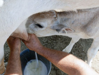
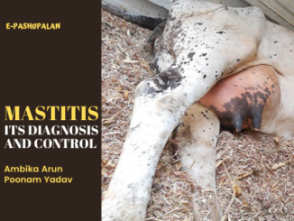
Incisional complications can be quite common following cesarean section. If necessary, some of the skin sutures can be removed so that the area can drain. Another common problem is retained placenta. Most cattle will “clean” or deliver their placenta after 24 hours. Antibiotics are often given until the placenta has been passed. The cow should be watched for dehydration and mastitis (mammary gland infection) after a cesarean section.
Prognosis and Prevention
Prognosis for recovery after cesarean section is generally favorable. To avoid C-sections, the cow and bull being bred should be of similar size and cows should be monitored carefully when they are close to delivery.
| The content of the articles are accurate and true to the best of the author’s knowledge. It is not meant to substitute for diagnosis, prognosis, treatment, prescription, or formal and individualized advice from a veterinary medical professional. Animals exhibiting signs and symptoms of distress should be seen by a veterinarian immediately. |

Be the first to comment