Abstract
Paratuberculosis, also known as Johne’s disease, is a chronic, wasting, granulomatous disease that affects ruminants, including yaks, and causes changes in antibody levels, a decline in animal production, and eventually death. The lack of diagnostic modalities to identify the diseases susceptible to yak during the early stages of infection creates significant barriers to disease control. The organism has become increasingly important in public health over the last decade due to two important factors. First, because of its increased evidence in animals, and second, because of its potential to enter the food chain, causing Crohn’s disease a debilitating condition. The aim of this paper is to determine the transmission routes, pathophysiology, diagnosis, treatment, and public health implications of Johne’s disease in yaks.
Introduction
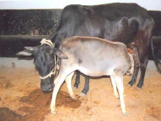
Mycobacterium avium ssp. paratuberculosis is the causative agent (MAP). Mycobacterium avium ssp. paratuberculosis is one of 24 species of Mycobacterium avium, some of which are pathogenic to humans and animals. The pathogen is a gram-positive, acid-fast, facultative intracellular pathogen that relies on iron for growth and can survive within macrophages. The bacteria is fastidious, and it causes disease in both animals and humans. The bacteria colonizes the yak’s intestine, causing production-limiting malabsorptive diarrhea, intestine dysfunction, and weight loss. Since the bacteria has a long incubation period, colonization of the intestine by MAP may take many years until overt signs of disease appear (between 2 and 7 years). As this disease is incurable, it worsens over time, causing emaciation and ultimately death of the infected animal. The yaks infected with JD remain asymptomatic for several years after contracting the disease. The chronology has been divided into four stages: silent, sub-clinical, clinical, and advanced.

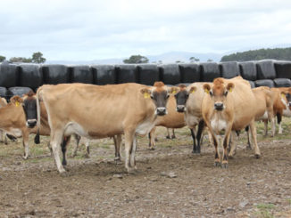
Yaks with disease stages ranging from subclinical to clinical continuously shed the organism in their milk and feces, contaminating the environment, pastures, and other members in the long run. The death of animals in JD or the culling of animals infected by the disease results in massive losses and economic uncertainty.
Transmission
MAP, the causative agent of JD, is transmitted through feces and milk of an infected yak and from mother to fetus across the placenta. As the clinical signs remain covert for several years after infection, the contamination of the milking parlor by the infected animals serves as one of the most common sources of contaminating milk with the bacteria. Human beings can also become infected by drinking contaminated milk and other dairy products.
Pathophysiology
Following ingestion of infected material, MAP bacteria enter the intestine and colonize Peyer’s patches by penetrating the mucosa with the aid of M cells (specialized mucosal cells for absorption) specifically at the ileum. MAP bacteria are phagocytized by macrophages but since they are immune to intracellular digestion, they are not phagocytized. There are no signs of infection in the animal at this time, which contributes to the low sensitivity of MAP diagnostic serological assays. Macrophages then spread the infection to regional lymph nodes via lymphatics, where the MAP bacteria cause a series of immunological and inflammatory reactions causing the body to establish a cellular immune response (primarily T lymphocytes) and produce inflammatory mediators’ viz. IFN-.y. IFN-y has been found to play a significant role in MAP resistance. The initial cellular response stops the infection from spreading in certain animals, but they remain sub clinically infected for the rest of their lives. Animals develop a humoral immune response with a lot of shedding when the initial cellular response is inadequate. The mechanism by which diarrhea occurs is thought to be antigen-antibody reactions with a resulting rise in histamine levels. Whatever the macroscopic lesions of the JD are, they are almost all concentrated to the intestinal part (mainly ileum) of the digestive tract and these include:
- Corrugated appearance of intestine (pathognomonic).
- Draining mesenteric lymph nodes.
- Edema and thickening of intestine.
- Dilation and thickening of mesenteric and serosal lymphatic vessels.
Diagnosis
The specificity of culturing the MAP from infected yak’s faeces, blood or milk sample has been found to be 100 percent and the sensitivity of culture diagnosis being higher if the animal is in clinical phase of disease in comparison to the animal being in pre-clinical stage of the disease. Most commonly used media used for culturing of MAP are Herrold’s Egg Yolk Medium, Lowenstein-Jensen medium, Jorgensen’s or Middlebrook 7H10 or 7H11 slants with the addition of Mycobactin.
The serological diagnosis can also be done by the techniques like ELISA, Gamma interferon assay, Complement Fixation Test (CFT), Agar gel immunodiffusion. However, the outcome of these techniques depends upon the stage of disease on which they are being utilized.
Another basis for diagnosis of MAP is molecular genetics, however its sensitivity as diagnostic tool is low.
Treatment
The protocol followed for treating JD is not only expensive, but ineffective as well, in addition to the therapy being followed for extensive periods. The various drugs used are rifampin, isoniazid, clofazimine, and streptomycin. Any one of these drugs or a combination of them has been seen to be somewhat effective.
Public Health Significance
A commissioned report by the National Association for Colitis and Crohn’s disease (NACC) in UK from an expert review group into the evidence linking MAP and Crohn’s disease in 2003 (NACC 2003). This report concluded that both live and dead MAP were present in human food samples and the strongest evidence for this is in milk and DNA of MAP can be found in the bowel tissue of a proportion of patients with CD but also in lesser quantities in the bowel tissue of human beings having no lesions of CD. Naser et al (2000) have detected the presence of antibodies against MAP in human breast milk and in 2004; he isolated MAP by IS900 PCR amplification from the blood and intestines of Crohn’s patients. Similar, findings were reported by Abubakar et al (2008) 4 years later.
References
- Abubakar, I., Myhill, D., Aliyu, S.H., Hunter, P.R., 2008. Detection of Mycobacterium avium subspecies paratuberculosis from patients with Crohn’s disease using nucleic-acid-based techniques: a systematic review and meta-analysis. Inflamm. Bowel Dis. 14: 401–410.
- Naser, S.A., Sagramsingh, S.R., Naser, A.S. and Thanigachalam, S. 2014. Mycobacterium avium subspecies paratuberculosis causes Crohn’s disease in some inflammatory bowel disease patients. World J. Gastroenterol; 20(23):7403-7415.


Be the first to comment