Introduction
- Avian tuberculosis is a chronic wasting, contagious disease caused by Mycobacterium avium.
- It occurs throughout the world in a variety of domestic fowl, Chickens, ducks, geese & turkey, sparrow, pigeon, ostrich etc .
- Disease also hold zoonotic importance & infect mammals such as swine ,mink, rabbit and human .
- This disease also be called avian mycobacteriosis or avian TB .
- Organism are generally resistant to number of disinfectants and survive outside the body for many years (R. Koch, 1882)
- Mycobacterium avium is also related to avium– intracellulare and form MAC which causes disease in immunocompromised humans ( in AIDS ) or in humans having chronic lung diseases , this complex commonly found in fresh water , house hold dust & soil
Sub species
- avium subsp. avium – MAA
- avium subsp. paratuberculosis – MAP
- avium subsp. silvaticum – MAS
- avium subsp. hominissuis – MAH
Morphology
- Intracellular
- Rod shaped: 1-3µm
- Weakly Gram positive
- Acid Fast
- Non motile ,non spore forming
- Slow growing mycobacteria
Disease is Characterized by chronicity, persistency in flock once established, induce unthriftiness, decrease egg production & finally death.
Occurred in older age poultry (Caged & stressed bird).
Organism does not show tropism for any particular tissue.
Very high no. of tubercle bacilli excreted from ulcerated tuberculous lesions of the intestine in poultry create a constant source of virulent bacterium ,respiratory tract is also a potential source of infection if lesion occur in tracheal mucosa .
Carcasses of tuberculous fowl & dressed offal’s for food is also a source of spreading the AT.
Transmission
- Ingestion is the most common route for transmission.
- avium is not pathogenic in guinea pig & rats but causes infection in rabbits & mice .
- Healthy pigs & monkeys carry the organism in digestive tract so it is not always spread through contact with infected birds.
- Infected birds, contaminated droppings, equips, litter , pens, tubercle bacilli carried by attendants shoe .
Pathogenesis
- Capacity of avium to produce disease appears to be related to cell wall constituents, which are certain complex lipid such as cord factor (trihalosedimycolate), Sulphur containing glycolipids, strong acidic lipids (mycolic acid) .
- Cord factor increases virulence, influence immune responses, induce the formation of granulomas & inhibit tumor growth .
- Delayed type hypersensitivity (DTH) mediated by lymphocytes develops after exposure. Once activated macrophages demonstrate an increased capacity to kill intracellular avium.
- Lymphocytes releases lymphokines that attract, immobilize & activated blood borne mono nuclear cells at the site of bacilli & their products .
- TNF alone or in combination with IL2 disassociated with macrophage killing of M. aviumserovar1.
- DTH which develops contributes to tubercle formation & is partly responsible for cell mediated immunity in tuberculosis.
Clinical Signs
- Depression
- Increased thirst
- Respiratory distress
- Decreased egg production
- Unilateral lameness
- Jerky hopping gait
- Tuberculous arthritis
- Nodular masses
- Knife edged keel bone
- Mortality insignificant
Diagnosis
1. Gross lesions
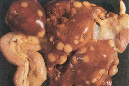
- Usually Seen in liver, spleen, intestine and bone marrow.
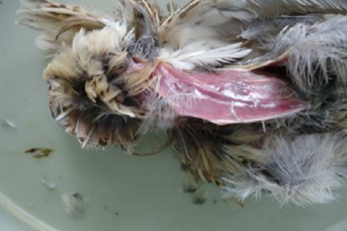
- Grossed emaciation with marked atrophy of sternal muscle & form prominent “knife edged keel bone”.
- Ulcerated lumen of intestine (primary site of lesions).
- Granulomatous lesion without calcification.
- Protrusion of tubercles from surface to the spleen gives it knobby appearance.
- Bone marrow of the long bones of legs (femero-tibiotarsal & tibiotarsal-tarsometatarsal joints) usually contains tubercular nodule (pale yellow in color , vary in size & numbers).


2. Microscopic lesion
- Giant cells are developed & arranged in palisade formation (fencing).
- Bacilli are more no. in central or necrotic zone of the tubercle & also very high no. in epitheloidzone.
- Final phase of tubercle formation is development of a zone of encapsulation.
- Caseous necrosis occur in central zone, calcification of tubercle occurs only & very rare in fowl.
- Acid fast bacilli occurs in great no. in smears of lesions.
3. Identification of the organism
Detection of acid fast bacilli in smears of infected liver, spleen, other organs. By ziehl nelson method or by auraminerhodamine florescent stain. (Magenta red colour to bacilli) Cultural examination for isolation & identification of organism on media lowenstin Jensen, herrold’s media, middlebrook 7h10 r 7h11, colitsos with 1% sodium pyruvate for 8 weeks .
- Multiplex PCR, Nucleic acid hybridization probes has a gold standard for distinction in avium & M. intercellular.
- RFLP – restriction fragment length polymorphism.
- Radiographs
- Cording/serpentine arrangement is absent in case of avian TB while it is a characteristic feature in Tuberculosis.
Conclusions
- No vaccine available.
- Test & slaughter policy applied in poultry farm.
- Treatment is very costly & long lasting.
- Due to risk of antibiotic resistance in humans, treatment of avian TB is not followed.
- Zoonotic risk for humans.
- significant threat in Assess the prevalence of potentially pathogenic mycobacteria.
- WHO listed M. avium as emerging pathogen.


Be the first to comment