An ancient disease with 100% fatal outcome. Discovered and named by Girolamo Fracastoro in 15th century.

Family: Rhabdoviridiae
Genus: Lyssavirus Type1
Size: Approx. 180 nm long and 75 nm wide with a single –strand, negative-sense RNA genome
Type: 1. Furious 2.Paralytic
Incubation period: Variable from 5 days to several years.
Diagnosis: Serum, CNS Fluid, Urine, Skin Sample, Corneal Smear, Brain Biopsy +PCR
Other Diagnosis: MRI
Wound toilet can reduce the chance of developing rabies upto 50%
Epidemiology
- Rabies is present in all continents except Australasia and Antarctica.
- Rabies virus doesn’t survive long outside the body.
- It is maintained either serial infection in wildlife or by cycle of infection in domestic dogs.
- Transmitted by bites of infected animal.
- Many different wildlife are vectors depending on geographic location.
- Vampire bats in south America
- Skunks in north America
- Mongoose in Africa
- Raccoons in united states recently
- All mammals are susceptible to rabies. These include pet animals, livestock, wild animals All though the red fox is the vector and reservoir of the disease, humans are mainly at risk from affected domestic animals such as cattle and cats .
- The disease occurs in more than 150 countries and territories. Worldwide, more than 55,000 people die of rabies every year.
Incubation Period
- The incubation period of rabies is quite variable in different cases. It is influenced by the amount of virus transmitted, virus strain, site of inoculation, host immunity, and the severity of bite.
- The incubation period is shorter in case the site of bite is near to the head and where the site has more nerves in comparison to the cases where the rabid animal inflicts a bite on the extremities. Similarly, deep and multiple wounds infected with higher concentration of rabies virus may result in a severe and rapid onset of disease.
- In dogs and cats, the incubation period may be around 10 days to 6 months; most cases become apparent between 2 weeks and 3 months.
- In most species, the incubation period in naturally occurring cases is about 3 weeks but varies from 2 weeks to several months.
- Experimentally, the average incubation period has been reported as 15 days in cattle, 10 days in sheep, and 12 days in horses.
Clinical signs in Dogs: Two forms in dogs
- Furious Form
- Dumb Form
Furious Form
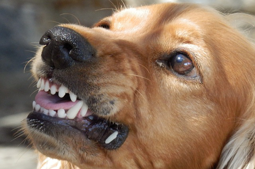
- Furious form of rabies is very common in dogs and the animal becomes quite excited and restless.
- A rabid dog becomes dangerously aggressive and bites objects like its own chain, stones, paper, wood, metal and even may show signs of snapping at imaginary objects. There may be unusual bark and aimless wandering.
- Later, there is drooling of saliva due to paralysis of muscles of deglutition. Partial paralysis of vocal cords leads to change in tone of bark to howl. The dog may not recognize it’s owner.
- In the terminal stage, there is muscular incoordination and paralysis of limbs and trunk. Death occurs mostly due to respiratory paralysis and convulsions.
Dumb Form
- Dumb form is the paralytic form of rabies which is characterized by progressive paralysis. In some cases it may develop without any noticeable signs of the furious form.
- Throat and masseter muscles become paralyzed and the animal may be unable to swallow. This causes saliva to accumulate with possible drooling. There may be facial paralysis.
- Dumb form of rabies is less common in dogs.
- The phase of excitement is short or absent in the dumb form. The dog has a dull or vacant expression and prefers to sit isolated in a corner.
- It may respond to its owner’s call but there is tendency of forgetfulness. Paralysis begins with the muscles of head and neck region.
- General paralysis results in death of the animal usually in 3–5 days.
Clinical Signs in Cats
- In a cat, the disease is usually of furious type.
- The cat may strike at the air with its forepaws as if it were catching mice.
- Paralysis of the hind part begins 2–4 days after the symptoms of excitement, and the animal generally dies in 3–5 days due to convulsions and respiratory paralysis.
Clinical Signs in Cattle
Furious form in cattle
- Cattle may appear unusually alert. Convulsions can occur, particularly in the terminal stages. In the final stages, there is incoordination and ascending paralysis.
- The animal usually dies 4–8 days after the onset of the clinical signs.
- Major clinical findings are excessive salivation, behavioral changes, muzzle tremors, bellowing, aggression, hyperesthesia, hyperexcitability and pharyngeal paralysis.
Paralytic Form of Rabies in cattle
- Early signs in the paralytic form of the disease include knuckling of the hind fetlocks, sagging and swaying of the hindquarters while walking and often deviation or flaccidity of the tail to one side.
- Drooling of saliva is one of the most constant findings. There are yawning movements which are more accurately described as voiceless attempts to bellow.
- When paralysis occurs, the animal becomes recumbent. Bulls in this stage often have paralysis of the penis. Death usually occurs 48 h after the recumbency develops and after a total course of 6–7 days.
Clinical Signs in Sheep and Goats
- Sexual excitement.
- Vigorous Wool pulling.
- Attacking behavior.
- Salivation
- Incoordination
- Muscle tremor.
- In one large outbreak deaths occurred between 17 and 111 days after exposure.
Signs of Rabies in Wild Animals
- Change in behavior of wild animals such as loss of fear of man or unusual friendliness.
- Nocturnal animals may show abnormal activity during daytime and may attack humans.
- Paralysis of the throat muscles can cause the animal to bark .
- Skunks, raccoons, and foxes usually display furious rabies. Bats often display dumb rabies. Unable to fly.
Manifestations in Humans :
- In humans, the incubation period is a few days to several years. Typically it is 1–3 months but may vary from less than 1 week to over a year. The length of the incubation period depends on factors such as the amount of virus inoculated, the degree of innervation at the site of viral entry, and the proximity of the bite to the central nervous system (CNS).
- The disease in the beginning appears with nonspecific symptoms. This stage is characterized with headache, restlessness, fever, and often pain or paraesthesia at the wound site.
- As the virus spreads through the CNS, progressive fatal encephalomyelitis develops and an encephalitic (furious) form or a paralytic (dumb) form may predominate.
- Neural symptoms of the disease include excessive salivation, excitation, aggression, behavior changes. Violent spasm of the gullet, pharynx, and larynx causes difficulty in swallowing of water and other fluids which is characterized by hydrophobia or aerophobia or both .
- The patient also becomes highly sensitive to noise and fearful of light (photophobia).These symptoms usually progress towards paralysis, coma, and death. In the terminal stages, there is respiratory paralysis, cardiac arrest and death.
Diagnosis in Animals
- In animals, rabies is diagnosed using the direct fluorescent antibody test (FAT), which looks for the presence of virus antigens in brain tissue.
Diagnosis in Humans
- Consequently, no tests are available to diagnose rabies infection in humans before the onset of clinical signs like hydrophobia or aerophobia.
- Tests are performed on samples of saliva, serum, spinal fluid, and skin biopsies of hair follicles at the nape of the neck.
- Saliva can be tested by virus isolation or reverse transcription followed by polymerase chain reaction (RT-PCR).
- Serum and spinal fluid are tested for antibodies to rabies virus. Skin biopsy specimens are examined for rabies antigen in the cutaneous nerves at the base of hair follicles.
Type of Exposure

Prevention and Control Strategies
- As dog is the principal reservoir of rabies, eliminating rabies in dogs is the most important approach for rabies prevention and control.
- Rabies prevention and control programmes have led to successful control or even elimination of rabies in several countries. Access to modern human vaccines and application of new economical post exposure prophylaxis (PEP).
- Animal birth control.
- Euthanasia of the rabid animals and street dogs.
- Identification and Recording.
- Proper disposal of carcasses.
- Vaccination of dogs and elimination of rabies in dogs constitute the mainstay of any rabies control programme.
- They should organize mass dog vaccination programmes in a planned manner.
- There is no evidence that removal of dogs alone has ever had a significant impact on dog population densities or the spread of rabies but targeted and humane removal of unvaccinated, ownerless dogs may be effective when used as a supplementary measure to mass vaccination .
Bite Wound Management
- The best way to ward off the exposure to rabies virus is to avoid bites and licking of fresh wounds or mucous membranes by the mammals. However, when such exposures take place, immediate medical care is important.
- For Category II and III exposures, this should be done by prompt and gentle thorough washing and flushing of the wound for about 15 min with soap or detergent and copious amount of running water. If soap and detergent are not immediately available, washing with running water alone is also useful. After thorough washing and drying the wound, disinfection of the wound should be carried out with ethanol (700ml/l) or iodine (tincture or aqueous solution), povidone iodine, or similarly viricidal topical preparation.
Pre-exposure Anti-rabies Vaccination
- Pre-exposure prophylactic vaccination is done to attain active immunity in high-risk individuals prior to a rabies exposure. It is recommended to those who are at continual, frequent, or increased risk of exposure to the rabies virus, either as a result of their residence or occupation. Such individuals include veterinarians, animal handlers, dogcatchers, wildlife wardens, quarantine officers, laboratory workers dealing with rabies virus or other lyssaviruses or infected material, postmen, delivery personnel, pet owners, and schoolchildren.
- Pre-exposure vaccination requires invariably three doses of intramuscular vaccination on days 0, 7, and 21 or 28.
Post-exposure Prophylactic (PEP) Immunization
- It should be initiated immediately following a transdermal bite or scratch by an animal suspected of being rabid or when possibly infectious material, usually saliva, comes into direct contact with the victim’s mucosa or with fresh skin wounds according to the WHO guidelines.
- Post-exposure anti-rabies immunization involves administration of anti-rabies vaccine with or without application of RIG based on the category of exposure as shown in Table .
- PEP treatment should be continued until a clear and unequivocal negative laboratory report is provided.
- All patients with Category II and III exposures require PEP vaccination.
- The 5-dose regimen: It consists of a 5-dose schedule. One dose of the vaccine is administered each on days 0, 3, 7, 14, and 28 intramuscularly in the deltoid muscle or anterolateral thigh muscle. This vaccination schedule involves five visits of the patient to the healthcare facility. It is also known as Essen regimen.
- The 4-dose regimen: This abbreviated multisite schedule, the 2-1-1 regimen, prescribes two doses on day 0, followed by one dose each on day 7 and day 21. On day 0, one dose is given in the deltoid muscle of the right arm and one dose in the left arm. Subsequently, one dose each is applied on day 7 and day 21 in the deltoid muscle. This vaccination schedule is also known as Zagreb regimen.
Dose of RIG
- The recommended dose of HRIG is 20 IU/kg bodyweight (total maximum 1,500 IU) and that of ERIG and F(ab´)2 products 40IU/kg bodyweight (total maximum 3,000 IU).
References:
- Veterinary Medicine –By Otto M Radostits.
- Textbook Of Preventive Veterinary Medicine- By Amalendu Chakrabarti.
|
The content of the articles are accurate and true to the best of the author’s knowledge. It is not meant to substitute for diagnosis, prognosis, treatment, prescription, or formal and individualized advice from a veterinary medical professional. Animals exhibiting signs and symptoms of distress should be seen by a veterinarian immediately. |


Thanks in support of sharing such a pleasant opinion,
article is nice, thats why i have read it fully
Good way of explaining, and pleasant article to take data concerning my presentation subject,
which i am going to convey in university.