Introduction
Canine parvovirus (CPV) still is and remains as a major canine pathogen in the world. Canine parvoviral enteritis has been a widespread and significant cause of morbidity and mortality in young dogs since its emergence in 1978. The virus’s tendency to “reinvent” itself and develop into new, more virulent, and resistant subspecies contributes to the virus’s continued occurrence of parvoviral enteritis.
Parvoviruses (Parvoviridae) are small, nonenveloped, single-stranded DNA viruses that are known to cause disease in a variety of mammalian species and that replicate in actively dividing cells single-stranded DNA virus that infects rapidly dividing cells of the gastrointestinal tract, bone marrow, lymphoid tissue, and cardiac myocytes1. Most parvoviruses are species-specific, hardy, surviving in the environment for long periods (5–7 months), and are widespread.
Epidemiology, Diseases transmission and Pathophysiology
Puppies between the ages of 6 weeks and 6 months are almost exclusively affected by this virus. The majority of adult dogs are resistant to the disease, either by natural infection or vaccination. If the bitch has antibodies to CPV, either from prior infection or vaccination, her puppies will be not be affected by parvovirus infection for the first few weeks of their lives. The amount of CPV-2 antibody transmitted to the neonate by an absorbed colostral antibody is 50–60% of the mother’s. The half-life of maternal parvovirus antibodies is about ten days.
Puppies become vulnerable to infection as their antibody titer decreases, and active immunization is necessary to protect them from disease. Lack of protective immunity, intestinal parasites, and overcrowded, unsanitary, and stressful environmental conditions are all factors that predispose puppies to parvoviral infection. Breeds like a rottweiler, Doberman pinscher, American pit bull terrier, Labrador retriever, and German shepherd dog have all been shown to be at an elevated risk of severe CPV enteritis.
CPV-2 spreads quickly among dogs through fecal-oral transmission (direct transmission) or oronasal exposure to feces-contaminated fomites (indirect transmission). The virus then replicates in the oropharyngeal and mesenteric lymph nodes, as well as the thymus, with infected animals being viremic in 1 to 5 days. Besides lung, spleen, liver, and kidneys, CPV-2 targets rapidly dividing cells of the intestinal epithelial crypts, bone marrow, epithelium of the tongue, oral cavity, and cardiac myocytes. Virus shedding typically occurs several days before clinical symptoms of vomiting and hemorrhagic diarrhea appear after exposure and an incubation period of 4 to 14 days. When enterocyte turnover is impaired, the intestinal lining becomes denuded, resulting in blunting of the intestinal villi, causing clinical symptoms such as vomiting and hemorrhagic diarrhea, as well as nutrient mal absorption and enteric bacterial translocation. The destruction and collapse of the thymic cortex are caused by a viral infection in the thymus. The virus causes the destruction of leukocyte precursors in the bone marrow, depletion of lymphoid tissues leading to significant leukopenia. If left untreated, the loss of immunity, combined with bacteremia from gut bacteria translocation, puts infected animals at risk of developing septic shock, systemic inflammatory response syndrome, multiorgan failure, and death.
Some factors especially parasitism and nonspecific factors (e.g., weaning), can increase mucosal cell activity, which may predispose dogs to clinical disease. This is usually attributed to changes in bacterial flora and diet during weaning, enterocytes in the intestinal crypts have a higher mitotic index and are therefore more vulnerable to viral tropism for rapidly multiplying cells.
Clinical findings
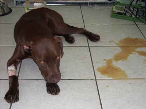
The gastrointestinal tract, bone marrow, and myocardium have all been associated with CPV infection, but the skin and nervous tissue may also be affected. The clinical response of dogs to CPV infection in the intestine varies greatly, ranging from undetectable infection to acute fatal disease. Anorexia, fatigue, depression, foul-smelling diarrhea that may range from mucoid to purely hemorrhagic, vomiting, dehydration, and fever are typical clinical symptoms of CPV-2 infection, which are nonspecific or referable to enteritis. In severe cases, elevated rectal temperature (40 ° to 41 ° C [104 ° to 105 ° F]) and leukopenia (primarily lymphopenia) may be present. Intussusception, a rare but potentially fatal complication of PVE, may occur due to intestinal dysmotility. Changes in mentation, prolonged capillary refill time, tachycardia, weak pulse quality/hypotension, cold extremities, and low rectal temperature are all symptoms correlated with tissue perfusion impairment. Puppies with underlying humoral immunity compromise, whether due to concomitant infection, low maternal antibody titers, or environmental stresses, have a more serious clinical disease. Some rare findings with CPV-2 have been documented including erythema multiforme, leukoencephalopathy, and porencephaly with periventricular encephalitis in puppies, as well as clinical disease and intracranial abscesses in cats.
The physical examination can reveal abdominal pain because of acute gastroenteritis or intussusception. Abdominal palpation, radiography, and ultrasonography can all be used to detect intestinal intussusception, which requires immediate surgical intervention.
Hematological and Biochemical Findings
Lymphopenia is the most common hematologic finding associated with CPV infection, but panleukopenia can also occur in severe cases. The hematological changes are widely accepted to be attributable to the destruction of hematopoietic progenitor cells of the various leukocyte types in the bone marrow and other lymphoproliferative organs such as the thymus, lymph nodes, and spleen. Other findings on a hemogram may include anemia (due to blood loss from GI tract as well as and due to rehydration therapy. and thrombocytopenia due to increased platelet use in the GI tract as well as caused by a reduction in platelet production or by viruses and/or immunologic components acting directly on platelets or endothelium.
Prerenal azotemia and elevations of hepatocellular enzymes due to extreme dehydration and tissue hypoperfusion; hypoalbuminemia secondary to gastrointestinal losses; hypokalemia secondary to gastrointestinal losses and inadequate intake; and hypoglycemia associated with severe malnutrition and/or underlying sepsis are all nonspecific abnormalities on serum chemistry evaluation. Other electrolyte imbalances (such as hyponatremia and hypochloremia) may also occur as a result of vomiting and diarrhea.
Diagnosis
Animals can shed CPV-2 virus in their feces within three days of infection, with peak shedding occurring four to seven days later. Fecal ELISA, Electron microscopy, viral isolation, fecal hemagglutination, latex agglutination, counter immuno electrophoresis, immunochromatography, and polymerase chain reaction are some of the other methods that can be used to detect CPV antigen in feces. Traditional techniques are less sensitive than PCR-based methods, especially real-time PCR.
Management and Treatment
In the absence of treatment, canine parvoviral enteritis has a less survival rate but with treatment, the rate rises to a higher percentage. Since there is no cure for CPV enteritis that is unique to an agent, the disease is managed by supportive care. Maintenance of hydration acts as core to supportive care because severe ongoing fluid losses resulting from vomiting and diarrhea are common in CPV enteritis. Intravenous crystalloids (e.g., lactated Ringer’s solution [LRS]. Nil per os for 24 to 72 hours has been recommended in the past for the treatment of CPV enteritis. Potassium may be supplemented depending on the serum potassium content of the particular patient. Hypoglycemia is a common occurrence. The concentration of glucose in the blood should be checked regularly and supplemented as required. If the serum glucose level falls below 60 mg/dL, an IV (or intraosseous) dextrose bolus (1–2 mL/kg 25 percent dextrose) should be given, followed by the addition of 2.5 percent to 5 percent dextrose to the crystalloid fluids (Normosol-R, Plasmalyte) are the preferred replacement fluids for the overwhelming majority of cases.
Maropitant (1 mg/kg subcutaneously every 24 hours) centrally acting antiemetic that acts via neurokinin-1 (NK-1) receptor antagonism. Metoclopramide, (0.5 mg/kg IV every 8 hours), a dopaminergic antagonist that blocks the chemoreceptor trigger zone and exerts a prokinetic effect in the upper intestinal tract, may be given as a bolus or as a constant-rate infusion in dogs with severe vomiting. Chlorpromazine (0.5mg/ kg intramuscularly or subcutaneously every 6 hours) is a phenothiazine derivative that blocks both the chemoreceptor trigger zone and the vomiting center in the brain. Ondansetron HCL, a 5-HT3 receptor antagonist that acts peripherally and centrally to inhibit vomiting, at a dose of 0.1–0.15mg/ kg intravenously every 6–12 hours, may be used in cases of intractable vomiting. Because of the destruction of the intestinal barrier and extreme leukopenia, puppies with CPV enteritis should be treated with intravenous, broad-spectrum, bactericidal antibiotics. A combination of a β-lactam antibiotic (ampicillin, 20 mg/kg IV every 8 hours) or a β -lactamase resistant penicillin (amoxicillin-clavulanate, 20 mg/kg IV every 8 hours) and an aminoglycoside (amikacin, 20 mg/kg IV, intramuscularly, or subcutaneously every 24 hours after the dog has been rehydrated; used for a maximum of 5 days). Puppies with CPV enteritis often have comorbidities, including gastrointestinal parasitism. With this in mind, antiparasite therapy should be initiated as soon as the puppy can tolerate oral therapies. Drugs like Pyrantel pamoate 5–10 mg/kg PO Q 24 h and Fenbendazole 50 mg/kg PO Q 24 h for 3–5 d can be used.
In patients with CPV enteritis, fluid loss in diarrheic feces, as well as a lack of enteral nutrition and the development of acute-phase proteins over albumin synthesis, can all lead to severe hypoproteinemia. A prophylactic administration of fresh frozen plasma (6.6–11 mL/kg IV or intraperitoneal, 3 doses 12 hours apart) can prevent serious infection in exposed dogs. The most commonly used synthetic colloids in veterinary medicine are hetastarch (20-30mg/kg/d) and dextran. Canine-specific albumin concentrate) is also available for administration and is a cost-effective way to restore serum albumin levels without the possibility of immune suppression associated with the use of concentrated human albumin products. Both provide a much greater oncotic pull per unit volume than the naturally occurring colloids yet remain relatively affordable and with a long shelf life.
The use of convalescent serum from dogs that have recovered from CPV infection (1.1–2.2 mL/kg intravenously or subcutaneously) as a passive immunization method. Oseltamivir is a neuraminidase inhibitor that has been researched in puppies with CPV enteritis. It is an antiviral drug used specifically for the treatment of human influenza. Granulocyte colony-stimulating factor (G-CSF) is a cytokine produced by bone marrow stromal cells, endothelial cells, macrophages/monocytes, and fibroblasts that causes granulocytes to be released from the bone marrow storage pool, shortened neutrophil maturation time, and increased granulopoiesis. Recombinant G-CSF (5 mg/kg) G-CSF (recombinant canine Granulocyte-Colony Stimulating Factor, provided once daily) increases white blood cell and neutrophil counts, as well as monocyte and lymphocyte counts, in a statistically significant way. The administration of probiotics to puppies with CPV increased clinical scoring in terms of dehydration, vomiting and diarrhea incidence, fecal scoring, and appetite. Transfaunation, or the administration of fecal transplants from a stable host to animals with acute hemorrhagic diarrhea, is another way of restoring fecal microbiota.
Prevention
Subclinical infection in wild and domestic dogs that shed viruses in their feces can be a significant source of infection for other dogs, particularly in crowded or unsanitary environments like shelters or breeding kennels. In crowded areas such as veterinary hospitals and shelters. Diluted 0.75 percent sodium hypochlorite solution applied to environmental surfaces is effective at substantially reducing the spread of CPV. Only isolating at-risk puppies from CPV exposure is the only way to avoid infection. Client education is critical in preventing at-risk puppies from being exposed to other pets before they have obtained their full course of vaccinations. Personnel in animal shelters and veterinary hospitals should wash their hands thoroughly after each patient and put on new gloves. Thermometers, stethoscopes, and other instruments, as well as clothing, instrumentation, chairs, cages, and bedding, should all be thoroughly washed and disinfected on a regular basis with appropriate detergent and viricidal solutions.
Vaccination
The most successful way of preventing CPV infection and disease, besides strict hygiene methods, is through careful and strategic inoculation with the production of protective antibodies. Parvovirus can infect dogs of any age or breed, but puppies between the ages of 6 and 16 weeks appear to be the most vulnerable. Vaccination with a high titer, low-passage, updated live vaccine should begin at 6 weeks of age and be repeated every 3 to 4 weeks until 16 weeks of age, as per current vaccination recommendations. Vaccination as early as 4 weeks to 18 to 20 weeks of age can be recommended for dogs with a substantially elevated risk of exposure (e.g., those in shelters). A booster vaccination is recommended at the age of one year and then every three years after that.
References
- Bonagura, J. D., & Twedt, D. C. (2008). Kirk’s current veterinary therapy XIV-E-Book. Elsevier Health Sciences
- Decaro, N., & Buonavoglia, C. (2008). An update on canine coronaviruses: viral evolution and pathobiology. Veterinary microbiology, 132(3-4), 221-234.
- Duffy, A., Dow, S., Ogilvie, G., Rao, S., & Hackett, T. (2010). Hematologic improvement in dogs with parvovirus infection treated with recombinant canine granulocyte‐colony stimulating factor. Journal of veterinary pharmacology and therapeutics, 33(4), 352-356.
- Goddard, A., & Leisewitz, A. L. (2010). Canine parvovirus. Veterinary Clinics: Small Animal Practice, 40(6), 1041-1053.
- Greene, C. E. (2006). Infectious diseases of the dog and cat (No. Ed. 3). WB Saunders\Elsevier Science.
- Mazzaferro, E. M. (2020). Update on Canine Parvoviral Enteritis. Veterinary Clinics: Small Animal Practice, 50(6), 1307-1325.
- Mischke, R., Barth, T., Wohlsein, P., Rohn, K., & Nolte, I. (2001). Effect of recombinant human granulocyte colony-stimulating factor (rh G-CSF) on leukocyte count and survival rate of dogs with parvoviral enteritis. Research in veterinary science, 70(3), 221-225.
- Mylonakis, M. E., Kalli, I., & Rallis, T. S. (2016). Canine parvoviral enteritis: an update on the clinical diagnosis, treatment, and prevention. Veterinary Medicine: Research and Reports, 7, 91.
- Prittie, J. (2004). Canine parvoviral enteritis: a review of diagnosis, management, and prevention. Journal of Veterinary Emergency and Critical Care, 14(3), 167-176.
| The content of the articles are accurate and true to the best of the author’s knowledge. It is not meant to substitute for diagnosis, prognosis, treatment, prescription, or formal and individualized advice from a veterinary medical professional. Animals exhibiting signs and symptoms of distress should be seen by a veterinarian immediately. |


1 Trackback / Pingback