Introduction
Heart worm infestation in dogs is a parasitic disease caused by a filarial organism, Dirofilaria immitis (Genchi et al., 2011). Mosquitoes (Aedes, Anopheles and Culex) can serve as intermediate hosts (Wang et al., 2014). Heart worm infection risk is greatest in dogs and most cases are diagnosed in medium to large sized, 3 to 8 years old dogs living outside. The classical symptoms of heartworm disease are coughing, exercise intolerance, unthriftiness, dyspnea, cyanosis, haemoptysis, syncope, epistaxis and ascites (Calvert, 2005). If the dog is not serologically tested, infection and disease progress remains undetected. Dogs show no indication of heartworm infection during the six-month prepatent period prior to the worms’ maturation and current diagnostic tests for the presence of microfilariae or antigens cannot detect prepatent infections.
The severity of cardiopulmonary pathology in dogs is determined by worm load, host immune response, duration of infection and host activity level. Live adult heartworm cause direct mechanical irritation to the intima and pulmonary arterial walls, leading to peri-vascular cuffing with inflammatory cells, including infiltration of high numbers of eosinophils. Live worms seem to have an immunosuppressive effect; however dead worms leads to immune reactions. Active dog tend to develop more pathology than inactive dogs for any given worm burden (Calvert, 2005).
Heartworm associated inflammatory mediators that induce immune responses in the lungs and kidneys cause vasoconstriction and possibly broncho-constriction. Canine heart worm disease type IV, also known as caval syndrome, is characterized by sudden onset with collapse. This syndrome is usually fatal; death may be due to acute respiratory distress and shock (McCall et al., 2008).
History and clinical observations
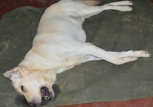
A Labrador male force sniffer dog, aged 5 years 8 months was presented with complaint of vomiting with yellowish discharge with normal rectal temperature (100.80F). As according to the history, animal was perfectly normal a day before and vomited after having meal. Contents of vomit were primarily of water and foam (common in gastritis). Animal was recently transferred to district Siddharthnagar (UP) from heavy mosquito infested area of Indo-Nepal border post.
Initially animal responded to treatment given for gastritis. Later in the evening animal vomited with presence of digested blood in vomit along with high rise of temperature (105.60F) which was not lowered down by antipyretics. On clinical examination dog was looking dull, depressed and apathic with mucous membrane pale pink, pulse- 72/min and CRT- ≥ 3 seconds and finally animal died in a short span of disease episode (12 hrs).
Treatment and discussion
On the basis of history of vomiting and clinical observation treatment for gastritis was given including Inj. Normal saline 150 ml Inj. RL 150ml IV, Inj. Metrogyl 50ml (250 mg) total dose IV, Inj. Intacef Tazo 562.5mg IV, Inj. Pheniramine maleate 1.5 ml IM, Inj. Stemetil 1ml IV, Inj. Pantaprazole 40 mg IV. Further it was advised to repeat the treatment after 12 hrs but in the evening temperature was 105.60 F. In that condition Inj. Melonex plus 1.5 ml IM was injected, however the condition of dog didn’t improved and animal expired same day.

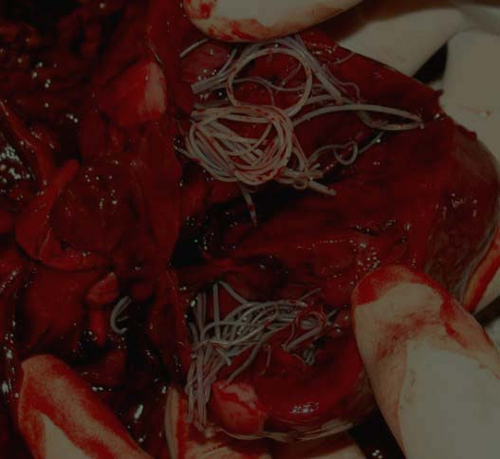
On post-mortem examination lesions in respiratory system including congested trachea, pleura and lungs were found, which indicated respiratory distress and right side congestive heart failure. Circulatory system exhibited congested pericardium, enlarged heart along with a bunch of heart worms. Enlarged and congested liver and spleen also expressed the picture of inappropriate circulation and CHF. Besides, congested intestine and blood mixed fluid was found in stomach.

Further visceral samples of dog were sent for histo-pathological examination. Histopathology report clearly revealed generalized inflammatory response of the body. In lungs it showed thickening of alveolar septa with infiltration of inflammatory cells, congested blood vessels along with cross section of parasite in the lumen of pulmonary artery, inflammation of lung parenchyma and inflammatory changes in bronchi. In kidneys, histopathology changes denote severe tubular necrosis and moderate atrophy of the glomeruli, which may be due to immune mediated inflammatory response. Histopathologic changes in stomach reflect acute gastritis. Splenic picture revealed severe depletion of white pulp and infiltration of inflammatory cells.

In view of clinical history, postmortem findings and histopathological examination it was suggested that the cause of death of animal may be renal- pulmonary dysfunction. However the finding of heartworms in the heart of the dog would be the primary cause which leads to changes in the vital organs. The death case of dog, classified as an acute failure of cardiac-pulmonary-renal system, which may be due to undiagnosed Dirofilariasis with sudden onset and collapse. This condition is fairly possible in absence of classical symptoms of heartworm disease i.e. coughing, exercise intolerance, unthriftiness, dyspnea, cyanosis, haemoptysis, syncope, epistaxis and ascites.
Heart worm is misnomer because the adult actually resides in the pulmonary arterial system and the primary insult to the health of the host is manifestation of damage to the pulmonary arteries and lungs (Ettinger and Feldman, 2010). Obstruction of pulmonary vessels by living worms is of little clinical significance unless worm burden is extremely high. The major effect on the pulmonary arteries is produced by worm induced (toxic substances, immunological response and trauma) villous myointimal proliferation, inflammation, pulmonary hypertension (PHT), disruption of vascular integrity and fibrosis. The condition further complicated by arterial obstruction and vaso constriction. Thus through this case, it may be concluded that in heartworm infestation it is fairly possible that a dog may collapse without revealing any classical symptoms.
References
- Calvert C. A. (2005). The Merck Veterinary Manual, 9th edition. Merck & Company, INC, White House Station, USA; 102.
- Ettinger, S. J. and Feldman, E. C. (2010). Textbook of Veterinary Internal Medicine, 7th edition. W.B. Saunders Company. ISBN 978-1-4160-6593-7.
- Genchi, C., Mortarino, M., Rinaldi, L., Cringoli, G., Traldi, G. and Genchi, M. (2011). Changing climate and changing vector-borne disease distribution: the example of Dirofilaria in Europe. Veterinary Parasitology; 176(4):295–9.
- McCall, J. W., Genchi, C., Kramer, L. H., Guerrero, J. and Venco, L. (2008). Heartworm disease in animals and humans. Advance Parasitology; 66:193–285.
- Wang, D., Bowman, D. D., Brown, H. E., Harrington, L. C., Kaufman, P. E. and McKay, T. (2014). Factors influencing U.S. canine heartworm (Dirofilaria immitis) prevalence. Parasite Vectors; 7:264.
|
The content of the articles are accurate and true to the best of the author’s knowledge. It is not meant to substitute for diagnosis, prognosis, treatment, prescription, or formal and individualized advice from a veterinary medical professional. Animals exhibiting signs and symptoms of distress should be seen by a veterinarian immediately. |


Glad to know from epashupala.com some clinics and dispensaries are having laboratory diagnostics. We have no such facilities in Arunachal Pradesh. We have long way to go to have sophisticated diagnostics and treatments.
Very good article presented by the author. It will be very useful for future reference.
Informative. Thanks to the author.
Nice presentation
Good informstion