Pneumonia is the inflammation of the pulmonary or lung parenchyma. It is usually accompanied by bronchitis and pleurisy. If pneumonia accompanied by bronchitis is called as bronchopneumonia, if it is accompanied by pleurisy is called as pleuro-pneumonia.
Predisposing factors for pneumonia
- Sudden change in climate means exposure to rainy or cold weather.
- Overexertion, hard work, prolonged strenuous exercise, long distance transportation.
- Malnutrition/ under nutrition, debility.
- Inhalation of dust, smoke, chemical vapors and faulty drenching of medications.
- Over crowded flock, poor ventilation, poor sanitation, excessive dust in animal shed.
- Cortico-steroid therapy, long standing antibiotic therapy, traumatic reticulo pericarditis.
Factors affecting incidence of pneumonia
- Age-Young and old one are more prone to pneumonia due improper immunity factor.
- Sex-Females are more prone to pneumonia compared to male animals.
- Status of immunity-immune compromised animals are more prone to pneumonia.
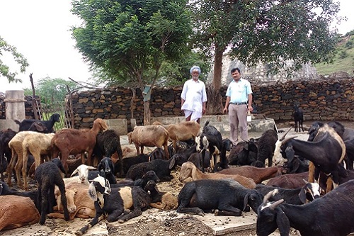
- Grazing animals- are more likely to get infection compared to stall fed animals.
- Stress- animal in stress are more prone to pneumonia.
- Bottle feeding/Weaning- During weaning period calf are more prone to infections including pneumonia.
- Dipping-improper dipping procedure and time will predisposes to pneumonia.
- Heavy worm infestation predisposes to pneumonia.
- Species – Cat are less susceptible to pneumonia.
Route of infection in pneumonia
- Bronchogenous route.
- Hematogenous route.
Classification of pneumonia
- Based on etiological agents which causes pneumonia.
a. Viral b. Bacterial c. Verminous d. fungal e. Aspiratory pneumonia. - Based on Anatomical part of lung affected.
a. Lobar pneumonia b. Bronchopneumonia c. interstitial pneumonia - Based on type of exudates in pneumonia.
a. Suppurative pneumonia b. Fibrinous pneumonia c. Pyogranulomatous pneumonia - Based on distribution of lesion on lung.
a. Focal pneumonia b .Lobar pneumonia c. Diffuse pneumonia - Based on Epidemiology of pneumonia.
a. Enzootic pneumonia b. Contagious caprine pleura-pneumonia c. Contagious bovine pleuro-pneumonia - Pneumonia in domestic animals classified into 4 types
a. Interstitial pneumonia b. Bronchopneumonia c. Aspiratory pneumonia d. Embolic pneumonia
Etiological agents causing pneumonia
Viruses will causes interstitial pneumonia and some of viruses causing pneumonia are
- In Cattle: BHV-1(IBR), Bovine respiratory syncytial virus, Parainfluenza -3 virus (Enzootic viral pneumonia– calves), Adenovirus 1, 2 and 3, Rhinovirus, Reovirus, BVD virus, Bovine mucosal disease complex virus.
- In Equines: Equine influenza virus, Equine Herpes virus 1 and 2, Alpha arteri virus(EAV), Equine Morbilli virus, Orbi virus(AHS).
- In Swine: Type A Swine influenza virus.
- In Sheep: Maedi(Progressive interstitial pneumonia), Jaagsiekte (Sheep pulmonary adenomatosis), Morbilli virus(PPR) Sheep pox virus, BT
- In Goat: Parainfluenza 3 virus, Reo virus, Morbilli virus(PPR),Goat pox virus, Pesti virus (Border disease)
- In Dog: Morbilli virus(CD), Canine influenza virus, Canine Herpes virus 1, Canine adenovirus type-2.
- In Cat: Feline Calci virus. Feline infectious peritonitis (FIP) virus, Herpes virus.
Bacteria will causes bronchopneumonia and some of bacteria causing pneumonia are
- In Cattle: Pasteurella multocida(HS),P hemolytica(Shipping fever), Klebsiella pneumoniae(calves), E.Coli, Salmonella, Pseudomonas, Sphareophorus necrophorus ,Streptococci, Staphylococci, Corynebacterium pyogenes, Mycobacterium tuberculosis (TB), Mycoplasma mycoides (CBPP) Actinobacillus lignieresi, M bovis, M avium.
- In Horse: Corynebacterium equi, Streptococcus equi, Pseudomonas mallei, E. Coli, Salmonella spp,, Pasteurella spp.
- In Sheep and Goat: P hemolytica, Klebsiella pneumoniae, E. Coli, Salmonella, Pseudomonas, Streptococcus spp, Staphylococcus spp, Mycoplasma mycoides(CCPP), Actinobacillus lignieresi.
- In swine: Haemophillus suis , Mycoplasma hyorhinis, Pasteurella multocida, Mycoplasma hyopneumoniae(Porcine enzootic pnwumonia), Bordetella bronchiseptica , Mycobacterium tuberculosis , Streptococcus spp, Staphylococcus spp.
- In Dog: Bordetella bronchiseptica , Klebsiella pneumoniae, Staphylococci, E. Coli, Salmonella spp,Pseudomonas spp. Streptococcus zooepidemicus, Pasteurella pneumonia, Mycobacterium tuberculosis, M bovis, M avium. Streptococcus canis.
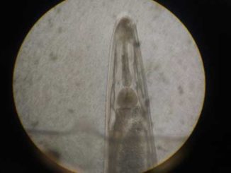
Worms causing pneumonia
- Cattle: Dictyocalus viviparus (Lung worm), Paragonimus westermani (Lung fluke) Ascarid larvae(Larval migration) Calves.
- Horse: Parascaris equorum (Larval stage migration), Dictyocalus arnfieldi (Obstructive bronchitis).
- Sheep and goat: Dictyocalus filaria(Lung worm), Mullerius capillarius (Nodular lung worm), Protostrongylus rufescens, Paragonimus westermani (Lung fluke).
- Swine: Metastrongylus apri, M salmi, M pudentodectus. Ascaris larvae(Larval migration).
- Dog: Angiostrongylus vasorum, Toxocara canis, Ancylostoma caninum, Toxoplasma gondii, Paragonimus westermani (Lung fluke).
- Cat: Alurostrongylus obstrusus (Lung worm).
Fungi (Mycotic pneumonia): Immunocompromised and super infection.
- Aspergillus fumigates (Brooder pneumonia), Histoplasma capsulatum, Blastomyces spp,.Cryptococcus spp, Mucor spp.
Foreign body Pneumonia: Common in ruminants due to penetrating type of foreign bodies to diaphragm then to lung.(TRP).
Aspiration pneumonia: Faulty drenching of drugs / liquids/mineral oils/liquid paraffin through oral cavity and paralysis/obstruction of larynx, pharynx and esophagus.
Embolic pneumonia: Ruminal acidosis, Liver abscess, mastitis, metritis, arthritis.
Stages of pneumonia
- Stage of congestion- Active hyperemia and edema in the alveoli. Lungs are congested and swollen. Microscopically-Capillaries in the alveolar wall are dilated and filled with blood.
- Stage of red hepatisation-Affected part will be cosolidated, red and looks like liver. Microscopically-alveoli revels fibrinous exudate having erythrocytes and polymorphonuclear leucocytes.
- Stage of grey hepatisation-Lung will be consolidated, looks like grey. Microscopically-ischemia of alveolar capillaries, thrombosis of alveolar capillaries, increased infiltration of leucocyte
- Stage of resolution-Liquefaction and removel of exudate. Lung backs to normal activity. Microscopically-Exudate will disappears, lung epiothelium regenerated. Sequelae or out come of pneumonia are death, atelectasis, abscess, gangrene, septicemia, incomplete resolution.
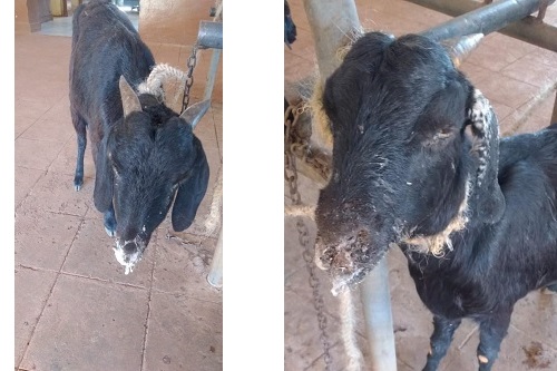
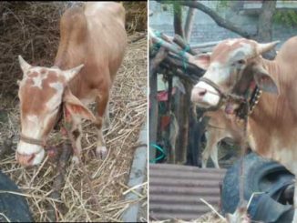
Clinical signs in pneumonia in animals
Fever, anorexia, dullness, depression, suspended rumination, fall in milk yield, tachycardia, difficulty in swallowing, running nose, falling behind in the flock, lethergic, weakness, noisy breathing. Respiration is rapid shallow breathing in early stage, dyspnea in later stage, coughing is moist, productive in bronchopneumonia (Bacterial origin), dry, unproductive in interstitial pneumonia(Viral origin), nasal discharges is bilateral serous followed by thick whitish discharge and bilateral ocular discharge. Animals will show Chest pain so they will reluctance to move and lie down and stand with abducted elbows with extension of head and neck. On percussion dull sound may be heard. On auscultation in early stage loud breath sound will be herd. Later stage – Moist/ dry rales observed In later stage consolidation will occurs shows increased audibility of heart sounds. Stunted growth, late maturity, rough hair coat, abortion, reduced feed and water intake, loss of body weight, high mortality in young ones, complications like nasal bots, maggot wounds, falling behind in the herd, self isolation from flock, prolonged calving interval.
Diagnosis of pneumonia
- History of change in climate, exposure to extreme climate, stress, Immunocompromise status.
- Clinical signs : Fever, dyspnoea, coughing, bilateral nasal discharge.
- Hematology :
- Leucocytosis with neutrophilia in bacterial infection.
- Leucopaenia in viral infection.
- Eosinophilia in verminous pneumonia.
- Radiographic examination: Chest X – ray increased radiopacity of lung in pneumonia, T. B. nodules, foreign body can be detected.
- Ultrasonographic examination: Normal lung-Hypoechoic, Consolidated n pneumonic lung-Hyperechoic.
- Isolation and identification of organisms-Nasal swabs, traheal swab/wash/ broncheo-alveolar lawage and staining of sample n culture of organism.
- Examination of faeces to detect larva stage L1 by Baerman technique.
- Serological tests like ELISA,HA HI test, PCR, Agar gel immuno diffusion test.
- CT Scan of lung, MRI Scaning of lungs, and thoracoscopy will helps in the diagnosis based on the lesions in the respiratory system.
- Bronchoscopy -Endoscopic examination of lung and bronchioles.
- Spirometry – Pulmonary function test.
- Blood gas analysis.
- Pleural fluid analysis.
- Sputum examination for Tuberculosis-Acid fast staining technique.
- Lung biopsy.
- Fluroscopy of lung.
- Necropsy findings-consolidated lung, hepatic changes, inflammatory exudate, haemorrhagic trachitis.
Therapeutic management for pneumonia
Specific treatment
- Use of broad-spectrum anti-bacterials/antibiotics.
- For Lung worm infestation: Broad spectrum anthelminthics
- Use of antifungal antibiotics for fungal origin.
- Non-steroidal anti-inflammatory agents as a supportive therapy.
- Anti-Histaminics
- Broncho-dilators
- Respiratory Stimulants
- Oxygen therapy
- Expectorants
- Nebulization therapy.
Prevention and control measures for pneumonia
- Avoid overcrowding of animals in the animal shed.
- Deworming schedule should be followed regularly.
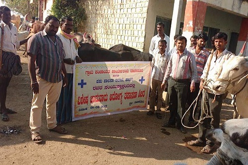
- Vaccination- PPR and HS vaccinations should be done at endemic areas.
- Avoid stress to animals.
- Avoid grazing during rain, extreme hot environment.
- Proper nutritious diet especially protein.
- Regular health monitoring in flock.
- Avoid corticosteroid therapy.
- Avoid late evening dipping.
- Proper hygiene and sanitation in the housing system.
- Special care to young ones
References
- Merck’s Veterinary Manual.
- Veterinary pathology. Ghanti, Shastri and A Ramarao
- MSD Veterinary manual
|
The content of the articles are accurate and true to the best of the author’s knowledge. It is not meant to substitute for diagnosis, prognosis, treatment, prescription, or formal and individualized advice from a veterinary medical professional. Animals exhibiting signs and symptoms of distress should be seen by a veterinarian immediately. |


Very nice 👌👌
Good information on pneumonia…