The third eyelid, also known as haws or nictitating membrane is a structure found in eyes of reptiles, birds and some mammals. It is a mobile, protective and glandular structure lying between cornea and lower eyelid in medial portion of the inferior conjuctival sac. It consists of a transparent membrane that is drawn across the eye and it serves a number of different functions. The third eyelid performs many important functions in dogs viz. protection of the cornea, distribution of tear film and production of immunoglobulins for the tear film. In fact, nictitating membrane acts like a windscreen wiper spreading the tear film evenly across the eyes in order to keep the cornea moist and healthy. These tears are produced by a special gland called as the nictitans gland, which is present at the base of third eyelid in the medial canthus of eye and is held in position by a ligament. The gland is innervated by sympathetic as well as parasympathetic nerves and blood supply is ensured by the nictitans arteries and veins. Third eyelid gland produces 30% of the total tears which are important for intactness of eyelid, eyeball surface and conjunctiva.
Cherry eye is a common term used for prolapse of third eyelid gland which is characterized by glandular swelling at the medial canthus of eye, hyperaemia and increase in the gland volume. It is a common condition encountered in canines. The eversion of nictitating gland is known as glandular hyperplasia, hypertrophy, nictitating gland adenoma, protrusion of gland or cherry eye. When the gland is protruded to outside environment, it makes the gland vulnerable to dust, dirt, microbes and wind leading to abrasion and desiccation of the gland. The gland gets hypertrophied due to secondary inflammation of the gland and conjuctival tissue in the surrounding. Thereafter, as the gland increases in size, it becomes difficult for it to return to its normal position. The medial canthus of the eye appears red and swollen due to this inflammation. It could occur in any age but most common in puppies. This can occur in 2-3 years of age and may be unilateral or bilateral. Breeds especially Pekingese, Neapolitan Mastiff, Cocker Spaniel, Beagle, Bulldog and Basset Hound are more prone to this pathological syndrome. The cause of the cherry eye is unclear. However, it is believed that it may occur secondary to inflammation and a weakness in the connective tissue attaching the gland to its normal position posterior and ventral to the nictitans, allowing the gland to migrate dorsally and flip up to protrude above the leading margin of the membrane. Pathogenesis of cherry eye has not been determined yet, but it may be associated with primary or secondary adenitis, fascial attachment abnormalities or some specific pathogens affecting the glands. Keratoconjuctivitis sicca may develop a few years later in the affected animals and it indicates that lacrimal glands may be involved along with nictitans glands. Cherry eye might not be a life threatening condition for pets but protrusion of the gland out of its normal position causes irritation to the animal along with giving a displeasing appearance to the owners with a pink, fleshy mass over the eye/eyes of their pets.
Diagnosis
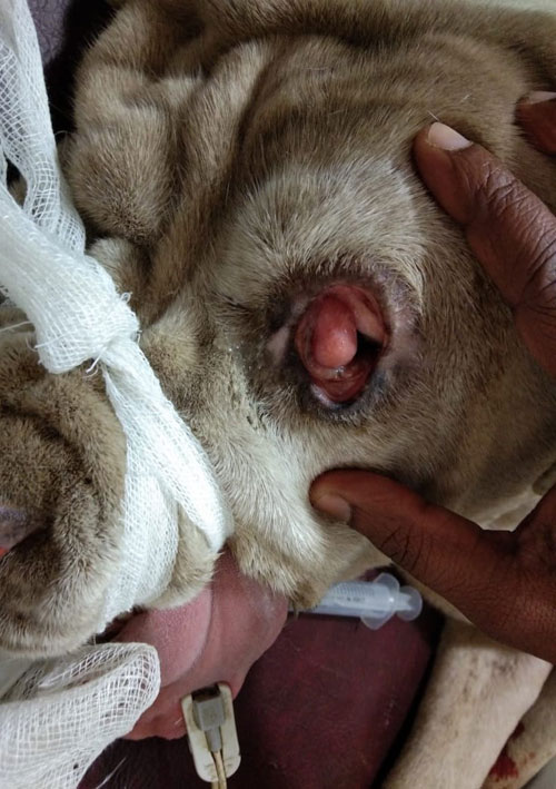
Diagnosis is done on the basis of history, physical and clinical examination. Generally during physical examination, the dog is found alert with a pinkish fleshy mass of tissue protruding from the medial canthus of left eye (Fig. 1).
Treatment
The management of cherry eye involves use of antibiotics, steroidal or non steroidal anti inflammatory ophthalmic preparations with a low success rate. Hence, surgical intervention is essential to reclaim normal condition of the animal. The two commonly practised methods of surgery are: Removal technique/ Excisional technique and Replacement technique. Both the above techniques have their own pros and cons. Earlier, removal of the gland was followed by surgeons, but the major concern in such cases is that the lacrimal gland might lose their function which renders the eye’s inability to produce enough tears for corneal protection. Hence, the removal technique is not in practice nowadays because of occurrence of dry eye condition which could further aggravate to Kerratoconjuctivitis sicca (KCS). This becomes a serious malady for the animal as KCS is an incurable condition. The only alternative in such a situation is regular administration of lubricants in eye and daily doses of lacrimogenic drugs, such as cyclosporine everyday for whole life of the pet. Failure to administer the daily doses will lead to pain and discomfort to the pet.
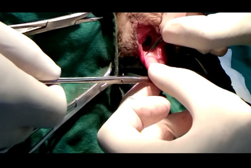
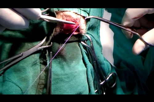
Replacement or pocket technique can be used to avoid the above condition (Fig 2). The pocket technique provides a good prognosis for permanent repair of prolapsed third eyelid glands. This procedure is helpful not only in alleviating the pain and discomfort of the animal but also helps in reducing the ill effects of surgery like reduced tear production. The suture material used is an absorbable suture to cause minimum irritation to the animal. The incisions are sutured and the gland is returned to its normal position using an absorbable multifilament suture material (Fig 3). Antibiotics are suggested for topical administration in the eye to reduce risk of further inflammation.
|
The content of the articles are accurate and true to the best of the author’s knowledge. It is not meant to substitute for diagnosis, prognosis, treatment, prescription, or formal and individualized advice from a veterinary medical professional. Animals exhibiting signs and symptoms of distress should be seen by a veterinarian immediately. |


Be the first to comment