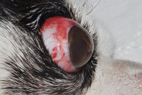
Traumatic Proptosis
Globe proptosis is a true ocular emergency. Proptosis is most often traumatic, the degree of trauma required to displace the globe varies greatly and depends on orbital conformation of the dog. Proptosis occurs more easily in brachycephalic breeds than in mesocephalic or dolicocephalic breeds. Immediate medical and surgical intervention is indicated to preserve the globe and vision, if possible.
Clinical observations
- Eyelids become trapped behind the equator of the globe due to trauma; this could result in impaired blinking and corneal exposure. Eyelid margins are seldom visible.
- Delay in the treatment could result in prolonged globe exposure that could increase the likelihood of corneal ulceration, retrobulbar oedema and haemorrhage and blindness from globe perforation or optic nerve damage.
- Enucleation is warranted when the globe is ruptured or more than two extraocular muscles or the optic nerve is avulsed.
Management of traumatic proptosis
- The cornea should be kept well-lubricated until globe replacement is achieved.
- Ocular surfaces are kept hydrated by using diluted povidone iodine (0.5%) with normal saline.
- Do not use povidone iodine scrub or chemical preparations (eg.) chlorhexidine to ocular surfaces which could cause corneoconjunctival ulceration.
- Administer systemic analgesics and apply topical lubricant (eg.) ophthalmic antibiotic eye ointment like chloramphenicol/chloromycetin/Gatifloxacin.
- Systemic corticosteroids (Prednisolone @ 0.5 -1.0 mg/kg body weight) should be administered immediately and continued at anti-inflammatory dosages for 7-10 days.
Surgical Measures-Temporary tarsorrhaphy
- Temporary tarsorrhaphy is placed to protect the globe from exposure.
- General anaesthesia should be induced as early as is safely possible, so that replacement of the globe can be performed.
Pre-operative preparations
- Ocular surfaces are cleaned using diluted povidone iodine (0.5%) with normal saline, eyelid margins and fornices are cleaned with sterile cotton tipped applicator with diluted povidone iodine (0.5%).
- Cornea, intraocular muscles integrity, vision, hyphema and pupillary light response (PLR) are checked to evaluate the globe’s condition to decide whether globe replacement or enucleation is indicated.
- Clip and prepare the eyelids and corneaconjunctival surfaces. Debris is removed from the cornea and conjunctiva with sterile saline or Ringer’s Lactate.
Surgical procedure
- A lateral canthotomy may be required to facilitate globe replacement, although an initial attempt may be made without performing a canthotomy.
- Full thickness (1 cm) lateral canthotomy is performed to lift the eyelid margins which are trapped behind the equator of the globe using tenotomy/ strabismus/ Mayo/ Metzenbaum scissors. Reposition the eyelid margins into their normal orientation facing forward and rostrally.
- The eyelid margins should be engaged with a strabismus hook or small retractor and everted while exerting gentle pressure on the globe with a lubricated gloved finger or flat side of a scalpel handle.
- Once the globe is replaced, 2 to 4 partial thickness horizontal mattress sutures are placed in the eyelids across the palpebral fissure to maintain globe position.
- Using stents like rubber band or intravenous tubing (iv) reduces skin tension from suture tension from the suture material. These stents prevent the pull-through and subsequent breakdown of the tarsorrhaphy and helps prevent pressure necrosis of the eyelids.
- Suture is first passed through the stent and then through the skin of the eyelid 5 mm from the eyelid margin directing it toward and exiting along the eyelid margin at the level of the meibomian gland openings. The suture material is left untied over the globe across the palpebral fissure. The needle is directed through the meibomian gland openings on the opposite lid, exiting the skin 5 mm from the eyelid margin and then entire procedure is reversed. Suture ends are tied after all have been placed.
- Canthotomy incision was closed with figure of eight suture to repose eyelid margins at the lateral canthus and prevent the suture material from protruding through the palpebral conjunctiva and irritating the cornea by placing the suture through the partial thickness of the eyelid. Exit the suture through the meibomian gland openings. Close the remaining canthotomy incision with simple interrupted suture.
- Use care that sutures enter and exit precisely at the eyelid’s leading margin. A small opening should be left at the medial canthus to facilitate topical medication.
- Tarsorrhaphy sutures can be left with long tags that can be tied in a bow and can be opened and closed as needed to apply medication or check’s the globe status.
Postoperative care
- Systemic broad spectrum anti-biotics, systemic anti-inflammatory medications (steroids) and analgesics for 3-5 days
- Topical broad spectrum anti-biotic ointments four times a day.
- collar is must to prevent the pre-mature suture removal by the dogs
- The tarsorrhaphy sutures should be left in place for a minimum of 10 days, at which time the most medial of the sutures may be removed. The remainder of the sutures may be removed 1 week later, in most cases.
Prognosis
- Factors which diminish prognosis include the presence of hyphema, secondary glaucoma, and avulsion of multiple extraocular muscles.
- Concurrent globe rupture, avulsion of all or most extraocular muscles or optic nerve avulsion are indications for enucleation rather than globe replacement.
- Good prognostic indicators include an obviously visual eye or one with an intact direct or consensual pupillary light response.
- Even eyes without a visual outcome may have a good cosmetic outcome, as long as there are no definite indications for enucleation. Due to avulsion of the medial rectus muscle, there will often be a residual lateral strabismus.
- Other complications may include keratitis (due to exposure, lagophthalomos, and/or corneal desensitization), KCS, and phthisis bulbi.
Conclusion
Proptosis is often traumatic and requires immediate medical and surgical interventions. The prognosis for vision is guarded to poor. Prompt treatment might save the globe.
Reference
- Plummer, C. E. Proptosis reduction. Clinician’s Brief. September 2013. Page no.67-71.
| The content of the articles are accurate and true to the best of the author’s knowledge. It is not meant to substitute for diagnosis, prognosis, treatment, prescription, or formal and individualized advice from a veterinary medical professional. Animals exhibiting signs and symptoms of distress should be seen by a veterinarian immediately. |


Be the first to comment