Introduction
The udder is responsible for milk production and delivery of milk and milk is an important source of income for the farmers. The quantity and quality of milk produced by the animals are directly affected by various disease conditions of the teat and udder. Cow and buffalo udder has four quarters each of which is a separate unit. Each quarter is an independent compartment. Affection of one quarter does not necessitate the involvement of the other quarters. In buffaloes the anterior teats are shorter than the posterior. In cow, the teats of the anterior quarters are longer than the posterior. This makes the anterior teats of the cow and the posterior teats of buffaloes more prone to injuries. Milk flow disorders due to traumatic teat injuries, faulty techniques of milking, infection, mucosal lesions, foreign bodies, milk stones and congenital disorders affect the udder health and milk production. This leads to a 50% increased chance of mastitis, which consequently leads to reduced milk yield and milk quality. Thus, early diagnosis and treatment of diseases of teat and udder is very important for maintenance of their health. The conventional diagnostic module includes inspection, palpation, probing, and hand milking of the affected teat, ultrasonography and contrast radiography.
Congenital and acquired surgical conditions of udder and teats can be grouped into three main categories.
- Conditions of epithelial surface of udder and teats.
- Conditions of Udder and teat canal.
- Conditions of teat sphincter.
For teat surgery, anesthesia can be obtained by a ring block at the base of the teat or local infiltration anesthesia in between the teats or inside the teat canal using 2 % solution of lignocaine HCl. The entire area is prepared for aseptic surgery by washing the field of the operation with antiseptic solution and water, and swabbing with alcohol.
- Conditions of epithelial surface of udder and teat
- Supernumerary or extra teats: This may occur and can be present anywhere on the udder but are most frequently seen posterior to the last two normally placed teats. These additional number teats may or may not have adjacent glandular tissue that will become functional. If there is a glandular tissue that has a functional potential, it will atrophy if not milked. For the management of this condition, it is better to amputate the accessory teats when that animal is young heifer, before the gland becomes active. It is essential that care must be taken to assure that only the supernumerary teats are removed and not normal. It may be desirable to remove the supernumerary teats for cosmetic reasons or because some may be so close to normally placed teats that they interfere with milking procedures.
- Fused teats: This may occur and can be present in between two collateral teats or the animals often has closely spaced teats on the opposite side. These Fused teats interfere with milking process which cause the milk splashes out. For treatment of fused teats, the skin is divided in between the two fused teats and cutaneous wounds are sutured separately.
- Udder and teat abscess: Abscess formation occurs more often on the udder than the teat. Many cases with chronic mastitis especially due to resistant microbes suddenly develop abscessation on side of affected udder. Such cases can easily be diagnosed by puncturing the swollen part. The abscess cavity is opened for complete drainage of pus under strict aseptic condition. After drainage of the pus, the cavity is dressed with tincture iodine followed by application of soothing agents until obliteration of abscess cavity. In case of necrosis of teat or udder, amputation of teat or affected quarter is recommended followed by daily dressing till complete healing of wound occurs.
- Sore teats: The teats become painful due to presence of cracks, traumatic injuries, lesions due to disease conditions such as pox, FMD etc. If these lesions are not treated well in time, the animal will not allow touching the affected teat for milking. In such cases, sterilized teat siphon should be used to drain the milk out. For treatment of such painful lesions, the wound should be washed with light potassium permanganate solution and then soothing preparation such as iodized glycerin, bismuth iodoform paraffin paste, zinc oxide ointment or antiseptic dressing with soothing emollient may be continued till the complete healing of the lesion occurs.
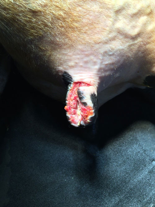
- Teat laceration: Lacerations of teat may be superficial or deep. Superficial skin lacerations of teats and or udder are treated with thorough cleaning and debridement of dead tissues and antiseptic dressing. In large wounds that do not penetrate sufficiently to allow milk to flow from the wound but muscles and skin are forming flaps, suturing should be done using a non-absorbable suture material after thorough cleaning and debridement of non-viable tissues. Deep lacerations involving the mucosa should be debrided first and a sterilized teat siphon is inserted. The mucosa is closed with simple interrupted or simple continuous pattern using absorbable suture. The submucosa and muscularis are sutured in simple continuous pattern. The skin is closed with simple interrupted or vertical mattress pattern using non-absorbable suture. Antibiotics are infused into the teat after surgery.

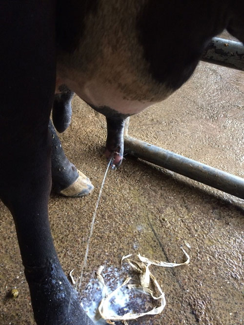
Teat laceration - Teat Fistula: The term, teat fistula, refers to an opening in the wall of the teat, connecting the exterior to the pre-existing channel, the teat canal is characterized by persistent outflow of milk. Such fistula may be congenital or acquired. It is mostly acquired as a result of penetrating wound that extend to the teat canal or cistern and fails to heal completely because of the continuous drainage of milk. Fistula will vary in size from that one which is so tiny, it is difficult to locate to large ones through which the mucous membrane may be seen. For the management of teat fistula, apply a suitable tourniquet (rubber band) at the base of the teat and a teat siphon to the affected teat. The wound edges should be debrided and flushed properly with antibiotic. Suturing of the teat fistulas is carried out in two rows including all layers with the exception of the mucosa using non-absorbable, non-capillary suturing material. A vertical mattress or similar stitch is used to effect the apposition of the edges deep in the tissue and superficially. The apposition must be complete and firmly held in place or milk seepage will cause the fistula to recur. A teat bougie is applied to prevent adhesion of both sides of the teat cistern and the tourniquet is then removed. The stitches may be removed in 10-14 days post operatively. Intra-mammary infusion of broad spectrum antibiotic for a week is indicated.

Teat fistula
- Conditions of Udder and teat canal
- Lactolith (milk stone): Milk stones which are found in the udder may result from accumulation of lime salts of milk over a point of crystallization. The latter may be desquamated epithelium. Sometimes, these calculi are freely movable in the teat canal if their sizes relatively smaller than the diameter of the canal. When being larger in size, they obstruct the lumen of the teat canal. If the calculi are of small size, they can be removed by manipulation during milking. Larger calculi obstructing the teat canal can be crushed by means of special forceps. In other cases of milk stones, it may be necessary to enlarge the opening at the end of the teat by cutting through the sphincter of the teat canal one or more times.
- Teat spider: This condition is usually due to congenital absence of teat cistern or canal. It can be acquired in cases of injury, tumour or inflammation of mammary tissue resulting in formation of thin or thick membrane, situated either at the base or middle of the teat. This membranous obstruction is removed using teat scissor, Huges teat tumour extractor, teat bistouries or Hudson spiral teat instrument.
- Fibrosis of teat canal: This condition is commonly observed in most of the lactating animals where a hard fibrous cord like structure is observed in the teat. Exact cause of this condition is not clear. However, repeated trauma due to mechanical injuries, thumb milking and calf suckling are the main contributory factors. Sometimes mastitis can also result into fibrosis of quarter followed by teat canal. This fibrotic cord will obstruct the teat canal and will create hindrance during milking. In such cases, initially hot water fomentation followed by counter irritant massage such as iodine ointment and turpentine liniment is very useful. In some cases it is advisable to place polythene catheter after removal of fibroid mass by Huges teat tumour extractor.
- Teat canal polyp: These are small pea sized growths attached to the wall of teat canal. The polyps hinder the milking process and sometimes even block the passage of teat canal. Teat polyps can be easily taken out by Huges teat tumour extractor. If its location is above the teat canal, thelotomy is the best method for resection of excessive tissue. Postoperative gentamicine and prednisolone infusion for five consecutive days found suitable to check infection as well as helpful in checking further growth of the polyp.
- Haematoma of the Udder: Haematoma of the udder is relatively common in cattle having pendulous udder as a result of contusion and rupture of a subcutaneous blood vessels. The condition is characterized by its sudden onset and fluctuency. Aseptic puncturing the swelling may be necessary to confirm diagnosis, but this is not preferable. If the haematoma is subcutaneous, it can be palpated out, if parenchymatus, it cannot be detected by visual examination and the diagnosis in such cases depends upon the sudden onset of bloody milk. Haematoma of the udder should be opened within a week post occurrence. The blood clot is removed and the cavity is painted with tincture of iodine. The cavity is then packed tightly to guard against further bleeding.
- Abscess of the Udder: Abscesses of the udder may develop beneath the skin as a result of infection of a haematoma. It may occur in the parenchyma of the udder as a result of chronic mastitis especially in goats. It may also occur as a result of supramammary lymphadenitis. Generally, abscess formations most commonly occurs secondary to the traumatic wound. Udder abscesses should be treated on the general principles for treatment of abscesses. If there are multiple abscesses, mastectomy (partial or total) according to the involvement of one quarter or more on the entire udder, is then indicated.
- Conditions of teat sphincter
- Contracted sphincter or teat orifice “hard milker”: The condition may be congenital in origin or may be acquired as a result of trauma to the end of the teat. There is a small stream of milk, and prolonged milking time. There may be loss of milk due to incomplete milking or trauma to the teat due to attempts for strenuous milking methods. Local infiltration anesthesia or instillation of 5 ml of 2 % Lignocaine or similar local anesthetic into the teat canal will provide anesthesia. The orifice should be cleansed, antiseptic applied, and the orifice enlarged. The enlarging procedure may be accomplished by inserting of lichty teat knife, ringed teat slitter or stoll teat bistoury. The opening in the sphincter is maintained at the desired size by inserting a Larson teat tube and leaving it in place for 5-7 days. Milking is accomplished by removing the cap of the tube.
- Enlarged teat orifice “Free Milker” or (Leaker): This condition is due to a relaxed or a traumatized sphincter. Milk leaks from the teat at times other than milking and result in milk loss. The condition may be helped by injecting small amounts of sterile mineral oil or lugol’s solution around the orifice to reduce its size to the desired effect. This may have to be done more than once to obtain the optimal size for milk flow. If it is overcorrected and result in stenosis, handle as contracted sphincter or orifice.
- Occlusion of the teat orifice “Blind teats”: This condition may be congenital or acquired due to any trauma near the teat sphincter. Such cases generally reported just after parturition. On palpation, milk thrill is found in teat cistern and on pressing, milk passed backward toward the udder cistern. Imperforated teat is treated by 15 gauge needle, after creating opening, it is further dilated using huges teat tumour extractor, milk canula fixed for 24 hours, after that frequent milking advised at 4 to 6 hours interval to prevent adhesion. Administration of proper antibiotics is adviced for a minimum period of 3-5 days.
If an animal is suspected for any of these surgical affections of udder treatment should be initiated immediately. Delay in treatment may worsen the surgical condition and may cause poor prognosis and leads to complications which may include occurrence of acute mastitis, increased somatic cell count, reduced milk flow and wound dehiscence. Postoperative care is very important for all these surgical conditions.
References
- Singh J, Singh P and Amold JP. 1993. The mammary glands. In: Ruminant Surgery (eds. Tayagi RPS and Singh J). CBS Publishers and Distributers, New Delhi, India, PP: 167 – 174.
- Purohit GN, Mitesh Gaur, Chandra Shekher. 2014. Mammary gland pathologies in the parturient buffalo. Asian Pacific Journal of Reproduction 3(4): 322-336.
- Horney FD. 1984. Bovine skin and mammary gland. In: Jennings PB, eds. The Practice of Large Animal Surgery. Philidelphia: WB Saunders PP: 263-271.
- Seeh C, Melle T, Medl M, Hospes R. 1998. Systematic classification of milk flow obstruction in cattle using endoscopic findings with special consideration of hidden teat injuries. Tierarztl Prax Ausg Grosstiere Nutztiere 26(4): 174-86.
- Singh P, Singh J, Sharma PD. 2003. Surgical conditions of udder and teats in buffaloes. Intas Polivet 4(II): 362-365.
- Rambabu K, Makkena S, Suresh Kumar RV, Rao TS. 2011. Incidence of udder and teat affections in buffaloes. Tamilnadu J. Veterinary & Animal Sciences 7(6): 309-311.
- Tiwary R, Hoque M, Kumar B, Kumar P. 2005. Surgical condition of udder and teats in cows. The Indian Cow 25-27.


Be the first to comment