Commercial advancement in late 1970s
Objectives
- Acceleration and proliferation of genetic material (complete genome male +female, in AI only male)
- Fertilized embryos transported around the world
- Disease control
- Bio security programme
- Genetic salvage of valuable individuals
- Development of new lines / breeds
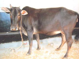
Donors selection
- Genetic merit
- Reproductive soundness
- Superior performance (individual or herd)
- Good BCS (preferably growing BCS)
- Free from diseases
- 50-60 days post partum
- Per rectal examination : cervix to ovary free with no adhesion
- Patency of cervical canal (esp Boss indicus) – should be patent
- Should have regular cyclicity
- Blood typing of both sires to further confirm offsprings (especially in cases of import of embryos)
Donor Management
- Selected from naturally cyclic group or those used for superovulation
- 2-4 donors for optimum efficacy
- 8-10 recipient for each donor
Superovulation (bovine)
- Aim: maximize the number of fertilized & transferable embryos
- Wide variation in superovulatory response
- Variability noticed for superovulatory response and embryo quality
- Dairy cows:
- Variability similar to beef
Variability in ovarian response due to
- Difference in superovulatory treatments
- Gonadotrophin preparation use
- Batch of gonadotrophin
- Dose of gonadotrophin
- Duration and timing of treatments
- Use of additional hormones
Animals and environmental factors
- Nutritional status
- Reproductive history
- Age
- Season
- Breed
- Effect of repeated stimulation
- Ovarian status at the time of treatment
- Environmental temperature /stresses
Gonadotrophins
Three preparations are used
Gonadotrophins (porcine or other animal pituitary) – eCG – hMG
- Pituitary extracts contain FSH
- Biological half-life of FSH in cow is 5 h or less
- Two injections daily (morning & evening) induces superovulatory response
- All injection by intramuscular route for 4-5 days
Doses
- Crude pituitary : 28-50 mg
- Partially purified (NIH-FSH-PI) : 260-400 mg (folltropin)
- Purified pituitary : 450 ug (porcine pituitary)
ECG
- Contain both FSH & LH has half life in cow is 40 h , persist up to 10 days
- Used as single injection I/M – PG after 48 h of eCG
Long half life resulted in
Continued ovarian stimulation and Unovulated follicles
Abnormal endocrine profiles and Reduce embryo quality
- Total dose (eCG) : 1500 -3000 IU (2500 IU commonly use)
- Intravenous administration of antibody to eCG 12-18h after onset of oestrus (time of AI) overcome some of the ovulatory problems
- Endocrine studies suggest that eCG treated animals had more frequently abnormal profile of LH and progesterone (reason for low production of transferable embryos)
- eCG produced from same mares had variations (FSH to LH ratio)
Experimental proof
Three groups exposed to pure FSH & different LH concentration
Results for better embryo quality & superovulatory response was with
group having lowest LH
- It was concluded that no additional LH is required for superovulation
- Endogenous LH is sufficient • Exogenous LH is detrimental to superovulation
- What is the best time for superovulation to optimize superovulatory response
Concept of follicular wave utilized – FSH used before the selection of DF
Experiment done using recombinant bFSH, treatment starts 1 day before, on the day, 1 or 2 day after wave emergence (based on the fact that endogenous FSH surge starts 1 day before emergence of wave)
Significantly more follicles were recruited when FSH treatments initiated on the day / day before wave emergence.
Traditional approach (followed for many years in ET)
- FSH treatment in mid cycle • Treatment of 4 or 5 days (no difference in ovulation)
- FSH used in decreasing or constant dose •
PG added on D 3 or D 4 of FSH treatment
- Mid cycle coincide with the period of second wave emergence
Drawback
- Skill person employed for oestrus detection (before FSH treatment and after FSH for insemination)
- All donors to undergo superovulation be in oestrus at the same time (need synchronization)
- Time wasted in initiating the superovulatory treatment (mid cycle waiting period)
Synchronizing wave emergence
- Use of E2 and progestins (1990s)
- E2 suppresses FSH release & follicle atresia
- After E2 metabolization, FSH surge & new wave emergence occurs
- On an average, wave emergence takes place 4 days after the E2 – progestin treatment
Oestradiol-17 β – 5 mg Or EB (oestradiol benzoate) – 2.5 mg (I/M)+P4 100 or 50 mg (I/M) & Progesterone implant (intra-vaginally) D 0 FSH treatments on D 4 (morning & evening)
PG on D 6 (M & E) – Removal of implant (D 6 evening)
Estrus on D 8 (48 h of PG) – AI (12 & 24 h after estrus)
- Disadvantage – Oestradiol banned in many countries
- Ablation of follicle
Used to suppress the effect of E2 on FSH and Procedure initiate new wave
Ultrasound guided oocyte aspiration used for ablation
5 mm or more diameter follicle aspirated
- Research further suggest to aspirate two large follicle (instead of all) to initiate wave
- Drawback
Required USG with pick-up facilities and trained person but Field application not possible
- Gonadotrophin releasing hormone (GnRH)
Used to induce ovulation of the DF & emergence of new wave 1-2 days later
Induction of ovulation is < 60% in random stage of estrus cycle
Lower superovulatory response compare to > follicle ablation
- How to improve ovulatory response
Pre-synchronization and – Synchronization of ovulation
Progestin insertion + PG administration (D 0) GnRH (7 days later for inducing ovulation) FSH treatment to start 36 h after GnRH
FSH treatment (4 or 5 days, if 5 days, remove progestin device a day later)
Remove progestin device (on D 4 FSH treatment )
- Note : • >95% animals ovulated to first GnRH
- Embryo number & quality were similar to oestradiol response (Reprodu Ferti Dev , 2010)
- Fixed time AI
Oestradiol + Progesteron device
FSH treatment (D 4) –PG ( M & E, D6)
Removal of progesterone (D 7,morning)
GnRH or LH (D 8 morning , 24 h after removal)
FTAI (12 h & 24 h later , i.e D 8 evening & D 9 morning)
Note
- Changes made by other workers – removal D 7 evening (additional 12 h) & GnRH or LH 24 h later (i.e D 8 evening)
How to avoid multiple use of FSH in superovulation
Drawback (multiple injection)
- Needs multiple handling of animals
- Painful to animals & some showed variations in preovulatory LH surge concentration
- Non-compliance of FSH schedule happens in certain cases
Single subcutaneous FSH injection
- Used in higher BCS beef cattle (> 3.0 on 5 scale) – Results were not repeatable in less BCS
- Superovulatory response similar to traditional
Mixing FSH with slow releasing polymers (hyaluronan)
- Tagged substance biodegradable & non-irritant to body tissue
- 2% hyaluronan solution used with FSH – Single injection FSH with 2% solution produces similar number of ova/embryo as of traditional method
Drawback
- 2% hyaluronan solution is much viscous and pose difficulties in FSH to dissolve
- Less viscous (diluted) concentration not effective as single injection
- Efficacy with less viscous solution was improved by splitting the dose (75% & 25%)
- All inj. given intramuscularly. Less concentration solution allows easy solubility of FSH
- In HF superovulatory response improved by splitting dose into two (75% & 25%)
- 75% S/C on first day – 25% 48 h (day of PG administration)
- Lyophilized FSH dissolved in 10 ml of 1% or 0.5% hyaluronan solution
- 75% dose on D 1 – 25% dose on D 3 (day of PG administration)
Embryo collection
- First successful non-surgical bovine embryo collection – 1976 by Dr Robert Rowe – Dr Peter Elsden– Dr Martin Drost
Time of Collection
- Calculation based on standing heat of donor
- Collection follows 6.5 day – 7.0 day
- Before or on D 6, embryo posses poor cryotolerance but D 7.5 – 8.0 emrbyos in hatched stage and becomes less useable.
- If multiple donors are used, flush the one who comes in heat first.
- Usually variation of 12-24 h in heat observed in multiple donor Superovulation
Collection media
- Commercially ready to use is best (not cost effective)
- Ringer lactate or D-PBS + 0.1% BSA, cheap and good for short term holding of emrbyos
- Embryos meant for export use polyvinyl alcohol (non animal resource)
Donor preparation
- Anteriorly elevated ramp (30-35 cm) to be placed in the chute (allow access to uterus easily as abdominal viscera shift posteriorly) – Donor should be full stomach (no need for fasting)
- Full stomach push uterus posteriorly, access to uterus easy.
- Empty stomach makes negative abdominal pressure, which causes air to sucked around arms and into the rectum and colon, making handling of uterus difficult
- When donor is in chute , give epidural anaesthesia (3-5 ml with 18 G needle)
- Tranquilization (10 mg xylazine) before epidural can be used for excitable animals
- Avoid using disinfectant at the site of epidural or perineal region cleaning.
- Simply wipe with water or clean towel the desired areas. Donor is ready for collection
Equipment
- Catheter : 52 cm, silicon, French foleys 16 /18G with 5 ml cuffs
- Stylet : 60 cm stainless steel
- ‘Y’ tubing : 150 cm plastic tubing, one end with syring tip and other for filter attachment
- Disposable three way plastic valves • 50-60 ml capacity air tight syringe
- Embryo filter 75µm • Jelly (non-spermicidal)
- Cervical dilator (ET / AI gun will be helpful)
- Flushing medium D-PBS +0.1% BSA • 10 ml syringe to inflate cuffs
- Sterilization of equipments
- Manufacturer supplied materials are gamma radiated sterilized
- Ethylene dioxide is embryotoxic
- Disposable items even can be reused (cost saving) by simply cleaning with lab reagent followed by multiple washing with distilled water
Embryo collection
- 52 cm silicon, 16/18 G French Foleys catheter is preferred even human Foley’s catheter 45 cm long , 16/18 G has been used
- Silicon catheter preferred as it causing minimum tissue irritation as compared to latex.
- The catheter is made rigid by inserting stylet.
- Stylet tip should be lubricate with jelly, which allow easy withdrawal
- Stylet should always be longer in length compare to catheter (52 cm vs 60 cm)
- After passing the stylet, catheter should be stretched to fit the stylet
- This procedure will prevent stylet to accidently pass into the fluid port
- In difficult cervix (‘s’ or 90º bending), cervical dilators (AI gun /ET gun) is helpful.
- Pass catheter immediately after dilator is removed otherwise cervix will collapse to its original shape • Squeezing cervical ring in front of the catheter will help in passing the catheter through tough cervix
- Opening of all cervical rings are not aligned and as such dilators is helpful
Body flushing
- Uterine body anatomy should be clear • Uterine body space varies 1.25 -5.0 cm
- Heifers body space 1.25 – 1.9 cm, aged animal 2.5 -5.0 cm
- Anterior to cuff, catheter tip length 2.5-3.75 cm
- Inflation of cuff will protrude the tip into the horn
- 2-3 ml inflation – 1.25 cm space occupied in the body
- 6-10 ml inflation – 2.5-3.75 cm space occupied
- Sufficient inflation required to place the catheter
- Inflation of cuff to be made with flushing media (any leakage can be detected at the valve , not so with air)
Placement of catheter into the body
- Placed catheter in horn (5.0 cm deep)
- Inflate with 1-2 ml fluid or till sensation of inflation perceived
- Retract stylet 5-6 cm into the catheter and than retract the catheter until cuff is in the body
- Continue inflating the cuff, tug catheter caudally to check sufficient inflation achieved
- Once inflation is sufficient, remove stylet
- Underinflated cuff pull back into the last cervical ring as the cervix relaxed
- Cervical pressure on the cuff block lumen of the catheter
Solution
Pinch behind the cuff in the cervix will squirt it back into the body. If this technique does not work, deflate, remove stylet, take out catheter and repositioned again
- Over inflation : the tip of the catheter will flush one horn
- After right placement of catheter, flush with media (gravity, syringe or pump) •
- Syringe infusion is preferred
- 400 ml media is required to flush both horns (50 ml x 8 = 400 ml)
- Before first flushing, the ‘Y’ tubing should be filled with media to remove trapped air.
- After each infusion, retrieved the maximum amount of infused fluid
- Half of the infused fluid come out by gravity, however, further retrieval required milking of the horns • Horn can be milked by grasping the tip of the horn between second and third finger and thumb used to pull the fingers (worm like movement)
- Gentle tug at the catheter on the body area will help in retrieval of fluid.
- With the last flush , infuse 25 mg PG into the uterine horn
- Before the use of embryo filters, separate evaluation in dish follows, which is time consuming. However, a beginner must use it.
- 400 ml fluid in 8 flush can be examined separately
- If embryos are detected in the lash dish (7-8) it indicates some embryos are left inside and need re-flush
- Also, after flushing the search in different dish, pass the fluid through embryo filter (75 μm ) in order to get any embryo which can not be searched
- Embryos of day 7 were present at the tip (Utero-tubal side) of the horn
- Heifers do not have a dilated apex (no pregnancy settled earlier) hence require more fluid to dilate and thus require more flushing media
Horn flushing
Take less time , Less media and Blockage of the oviduct can be detected
- Placement of catheter near 1/3 of apex (deep inside the horn)
- Cuff should not be overinflated (shape should be elongated and not round)
- Over inflation leads to rupture of endometrial wall and fluid leaked into uterine wall and broad ligament (crepitating sound)
- If rupture occurs, reposition the catheter ahead of the rupture site
- If under inflation, the catheter will slip
- Under such condition, re-inflate to go for body flushing


Be the first to comment