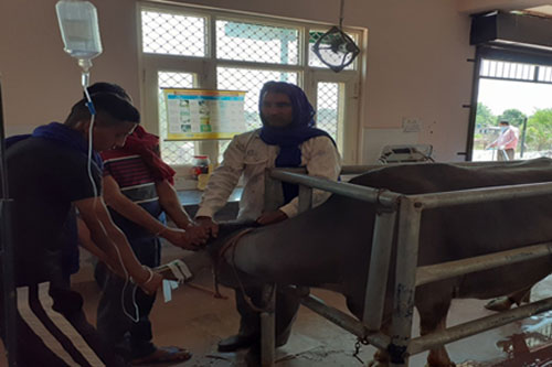
Introduction
About 70% of the body weight of the animals is made out of water. Water is very much needed for various vital activities such as metabolic processes; excretion of metabolic wastes; transport of enzymes and ions; maintenance of blood volume and blood pressure; thermoregulation; buffering agents and lubrication. About 50% of the body water is intracellular, rest is extracellular. The extracellular water is made of two fractions known as interstitial water which bathes the cells, and intravascular or plasma water. Of the extracellular water 25% remains as interstitial water and 5% as plasma water. The proportion of interstitial water is usually 3:1.
Indications
- Shock
- Severe and prolonged diarrhea or vomiting
- Anorexia due to infections, pyrexia, digestive disorders and postoperative
- Acute carbohydrate engorgement
- Abomasal displacement
- Intestinal obstruction
- Renal insufficiency
- Burns
- Copious sweating
- Lack of thirst or inability to drink water

The common forms of fluid and electrolyte imbalances are:
- Dehydration
- Electrolyte imbalances
- Acid-base imbalance
1. DEHYDRATION
Dehydration means loss of water and electrolytes from the body.
Causes: Dehydration is either due to failure of water intake or excessive loss of water.
- Failure of water intake
- Water deprivation
- Inability to drink due to painful conditions of mouth, pharyngitis, pharyngeal paralysis, oesophageal obstruction, coma.
- Lack of thirst due to toxemia or debility.
- Excess loss of water
- Gastroenteritis– vomition, diarrhea.
- Acute carbohydrate engorgement in ruminants.
- Abomasal displacement, impaction, pyloric stenosis.
- Intestinal obstruction.
- Severe peritonitis and ascites.
- Polyuria – Diabetes mellitus, Diabetes inspidus, Chronic renal failure.
- Burns and profuse sweating.
Clinical Signs
- Dryness and wrinkling of skin
- Sunken eyes
- Loss of body weight
- Muscular weakness
- Dry mucous membranes
- Oliguria
- Hypothermia – Cold skin and extremities
- Anorexia
Table 01: Assessment of degree of dehydration
|
S.N. |
Degree of dehydration (%) | Sunken eyes | Retention of skin fold (Sec) |
PCV (%) |
|
1. |
4-6 | – | Nil | 40-45 |
|
2. |
6-8 | Slight | 2-4 | 50 |
| 3. | 8-10 | Moderate | 6-10 |
55 |
| 4. | 10-12 | Deep | 20-45 |
60 |
Types of Dehydration
1. Isotonic dehydration
- Equal loss of water and electrolytes from the body.
- Dehydration usually moderate in nature.
- Causes : Simple enteritis, Profuse sweating, Nephrosis.
- Treatment : Normal saline or Ringer’s solution IV.
2. Hypertonic dehydration
- It means loss or deprivation of water with minor loss or deprivation of sodium.
- It is uncommon and dehydration is usually mild in nature.
- Causes : Water deprivation, Inability to drink water, Diabetes.
- Treatment : Water orally or dextrose 5% IV.
3. Hypotonic dehydration
- It occurs when there is loss of electrolytes in excess of water.
- Dehydration is severe / fatal in nature.
- Causes : Severe diarrhea (Salmonellosis / Colibacillosis), Intestinal obstruction.
- Treatment : Normal Saline, Ringer’s solution or Sodium chloride (5%) IV.
Basic Fluid Physiology
Fluid and electrolyte levels in the body are kept relatively constant by several homeostatic mechanisms. Normally, fluid is gained from animals’ food and drink intake (including a small amount from carbohydrate metabolism). It is lost via the urine, sweat and faeces, as well as through insensible losses via the lungs and skin.
![]()
Within the body, water is distributed into intracellular and extracellular compartments. The extracellular comprises both interstitial and plasma compartments. Water moves freely across the membranes that separate the compartments to maintain osmotic equilibrium.
![]()
Sodium-potassium pumps on cell membranes normally ensure that potassium is pumped into cells and sodium is pumped out, thus the intracellular sodium concentration is lower than the extracellular sodium concentration (the reverse applies to potassium).
![]()
In healthy animals’, volume homeostasis is regulated largely by antidiuretic hormone (ADH). Osmoreceptors and baroceptors detect small decreases in osmolality and blood pressure, triggering the release of ADH. This elicits a sensation of thirst and reduces renal excretion of water.
![]()
Renal mechanisms also play a part in volume homeostasis– the renin-angiotensin mechanism is activated by falling renal perfusion pressure.
It is important to remember that normal homeostatic mechanisms may not work well after injury (due to trauma or surgery), or during episodes of sepsis or other critical illness.
2. ELECTROLYTE IMBALANCES
- Electrolytes maintain osmotic pressure of body fluids and thereby maintain constant volume of different fluids.
- The electrolytes of major importance are sodium, potassium, chloride and bicarbonate.
- The electrolyte imbalances occur commonly as a result of loss of electrolytes, shifting of certain electrolytes or relative changes in concentration.
1. Hyponatraemia
- It means depletion of sodium ion.
- Sodium is the most abundant ion in extracellular fluid and is mainly responsible for maintenance of osmotic pressure of extracellular fluid.
- Causes : Colibacillosis, Salmonellosis, Acute diarrhea.
- Signs : Muscular weakness, mental depression, hypothermia, dehydration.
- Treatment : Normal saline or Hypertonic salt solution.
2. Hypokalemia
- Potassium is predominant ion in intracellular fluid.
- Hypokalemia means decrease in serum potassium level.
- Causes : Abomasal ulcers, Intestinal obstruction (High), Enteritis, Low dietary intake, Gastritis in dog. Prolonged used of mineral corticoids.
- Signs : Muscular weakness, recumbency, depression, cardiac arrhythmia, tachycardia.
- Treatment : Normal saline or Ringers’s solution or Isotonic saline + Potassium chloride (2g/lit.)
3. Hypochloraemia
- Hypochloraemia means decrease in chloride level.
- Causes : Abomasal disorders, Intestinal obstruction (High), Enteritis, Acute gastric dilatation in horse, gastritis in dog.
- Signs : Anorexia, weight loss, dullness, mild polydipsia and polyuria.
- Treatment : Normal saline or Ringer’s solution or Isotonic saline + Potassium chloride (2g/lit.)
3. ACID– BASE IMBALANCE
- The blood PH is maintained in the range of 7.35 – 7.45 by buffering system. The proportion of the components of buffer system viz. dissolved CO2 and bicarbonates are maintained at constant level by respiratory adjustment and urinary excretion of bicarbonate and hydrogen ions.
- The acid-base imbalance may lead to either acidosis or alkalosis.
Acidosis
It develops due to excess production of acid metabolites or as a result of loss of alkali (i.e. bicarbonates) from body or retention of CO2.
Causes
1. Metabolic acidosis: It is due to
- Excess loss of alkali i.e. bicarbonates – diarrhea.
- Increased production of acid in starvation, fever, shock, burns, diabetes mellitus, ketosis, acute carbohydrate engorgement in ruminants.
- Retention of phosphates, sulphates etc. in renal failure.
- Administration of excess quantities of acidifying solutions.
2. Respiratory acidosis: It develops when there is retention of CO2 in blood due to interference with normal respiratory exchange. It is commonly observed under following circumstances.
- Poor pulmonary ventilation : pneumonia, Pulmonary emphysema, Pleurisy, Congestive heart failure.
- Depression of respiratory centre due disease or drugs (anaesthesia).
Signs: Depression, muscular weakness, increased rate and depth of respiration.
Treatment: Ringer lactate solution or sodium bicarbonate orally or IV.
Alkalosis
It results from excess loss of acids or increased absorption of alkali or deficit of CO2.
Causes
1. Metabolic alkalosis: it may be due to
- Excessive loss of acids : Gastric dilatation in horse, Abomasal disorders and intestinal obstruction in ruminants.
- Overdosing with alkali viz : Bicarbonates.
- Excess absorption of alkali : Urea toxicity.
2. Respiratory alkalosis:
- It occurs in cases of hyperventilation resulting in excessive expulsion of CO2 and low blood carbonic acid.
- The common causes are high fever, toxaemia, encephalitis and brain tumour.
Signs : Slow, shallow respiration (to preserve CO2), muscle tremor and tetany.
Treatment : Normal saline, Ringer’s solution/Potassium chloride (1.1%) sol.
Table 2: Commonly used fluids : their composition and indications
|
Fluid and its composition |
Indications |
| 1. Normal saline (Sodium chloride 0.9%) | Expand circulating blood volume. |
| 2. Dextrose saline (Sodium chloride 0.9%, Dextrose 5%) | Severe sweating, Vomiting, Pyloric obstruction, Abomasal disorder |
| 3. Balanced Electrolyte Solutions | |
| a) Ringer’s solution (NaCl 0.9 g + KCl 0.03 g + CaCl2 0.03 g + D W 100 ml) | Dehydration, Vomition, Heat Stroke, Mild Diarrhoea |
| b) Lactated Ringer’s solution (As for Ringer’s solution + Sodium lactate) | Mild to moderate acidosis with dehydration. |
| c) Rintose/Intalyte 500 ml (Dextrose 20 g + NaCl 0.6 g + KCl 0.04 g + CaCl2 0.027 g + Na lactate 0.31 g) | Dehydration, Ketosis, Mild to moderate acidosis. |
| 4. Sodium bicarbonate solutions | |
| a) Sodium bicarbonate 1.3% (Isotonic) | Acidosis. |
| b) Sodium bicarbonate 5% (Hypertonic) | Severe acidosis. |
| 5. Mixture of isotonic KCl2 (1.1%) and Dextrose saline | Metabolic alkalosis. |
| 6. Dextrose Solutions | |
| a) Dextrose 5% (isotonic) | High Fever, Starvation, Decreased water intake. |
| b) Dextrose 20%, 25% (hypertonic) | Parenteral Nutrition, Ketosis, Hypogylycemia, Surra. |
| 7. Amino acid solution (Alamin SN, Hermin Amino drip 200, 500 ml) | Parenteral nutrition, Liver disorders, Hypoproteinemia, Extensive burns. |
| 8. Plasma Expanders | |
| a) Dextran-70, 500 ml (Dextran 6% in normal saline) Dose – 10 to 20 ml/kg/day IV | Shock, Haemorrhage, Burns, Severe dehydration, Surgical operations. |
| b) Haemacel, 500 ml (Polymer form degraded gelatin 3.5% + Electrolytes Na + K + Ca + Cl) | Hypovolemic shock, Haemorrhage, Burns, Endotoxic shock. |
| 9. Mannitol 20%, 100, 300 ml Dose @0.5-2 g/kg IV | Cerebral oedema, Meningitis, Encephalitis. |
Table 03: Fluid Therapy indicated in various diseases’
| Disease/Disorder | Fluid therapy suggested |
| 1. Enteritis (Diarrhoea) | Normal saline/Dextrose saline & Ringer’s solution/Sodium bicarbonate (1.3% or 5%) solution. |
| 2. a)Gastritis (Vomition), Abomasal Disorders, Intestinal obstruction in cattle, Alkaline indigestion | Normal Saline / DNS and Ringer’s solution / isotonic potassium chloride (1.1%) solution. |
| 3. Acid indigestion (Carbohydrate engorgement) | Sodium bicarbonate followed by Ringer’s solution. |
| 4. Stomatitis/Pharangitis/ Choke/Starvation/Anorexia/ Fever/Simple indigestion | Dextrose 5% or Dextrose Saline. |
| 5. Toxaemia: Black-leg/Botulism/ Enterotoxaemia/ Colibacillosis/ Salmonellosis | RL, Dextrose 5% and Ringer’s Solution. |
| 6. Heat stroke | Dextrose Saline. |
| 7. Acute renal failure (oligouric) | Dextrose 5% and Sodium bicarbonate. |
| 8. Chronic renal failure | Dextrose 5% and DNS in 2:1 proportion. |
| 9. Urolithiasis in bullock | Isotonic Saline and Potassium chloride (1.1%) along with calcium bicarbonate. |
| 10. Hepatitis | Dextrose 5%, 10% |
| 11. Ketosis | Dextrose 20% followed by sodium bicarbonate. |
| 12. Diabetes mellitus | Normal Saline. |
| 13. Diabetic ketoacidosis | Sodium bicarbonate 1.3% |
| 14. Milk fever | Calcium borogluconate. |
| 15. Metabolic haemoglobinuria | Sodium acid phosphate 20% sol. |
| 16. Burn, Haemorrhage, Shock, Severe diarrhea | Dextran – 70, Haemacel or Plasma. |
| 17. Cerebral oedema | Mannitol 10 & 20% |
| 18. Poisoning | Dextrose 5 & 10% |
ADMINISTRATION OF FLUID
1. Dose
The quantity of fluid required depends upon degree of dehydration, losses occurring during the treatment and maintenance requirement of animal.
The fluids are given in two stages –
1.1. Hydration therapy
- It is given in first 04-06 hours.
- It restores circulating blood volume and corrects electrolyte imbalance.
- Intravenous route is preferred for this therapy.
- The dose of fluid for hydration therapy is circulated as follows :
|
S. N. |
Dehydration | % |
Dose (ml/kg bw) |
|
1. |
Mild | 4-6 | 25 |
|
2. |
Moderate | 6-8 | 50 |
|
3. |
Severe | 8-10 | 75 |
|
4. |
Very severe | 10-12 | 100 |
1.2. Maintenance therapy
- It is given in next 24 hours (following hydration therapy).
- It is given for maintaining restored blood volume.
- It can be given by both intravenous and oral route.
- The maintenance dose is calculated @ 50 – 100 ml/kg body weight over period of 24 hours.
2. Route
The route of administration of fluid depends upon type of disease, condition of the patient, severity of dehydration and type of electrolyte/acid-base imbalance.
The fluids can be given by following routes.
2.1. Oral Route
- It is easiest route.
- It is preferred for maintenance therapy.
- It is unsatisfactory for hydration therapy.
- It is advocated in mild cases.
2.2. Intravenous Route
- It is preferred for hydration therapy and correction of electrolyte and acid-base imbalance.
- Hypotonic solutions are not used.
- Hypertonic solutions may be given.
- Large doses of fluids can be given.
- Fluid and electrolyte imbalances can be rapidly corrected.
2.3. Subcutaneous Rout
- Hypertonic solutions and plain glucose solutions are not given.
- Large amount of fluid can’t be administered.
- It is contraindicated in oedema.
2.4. Intraperitoneal Route
- It is preferred in large animal practice.
- Asepsis is important.
- Contraindicated in ascites.
3. Rate of administration
It depends upon following factors such as:
3.1. Size of animal: Fluids can be given at rapid rate in large sized animals.
3.2. Severity of dehydration: In severe cases, fluids are given rapidly.
3.3. Type of fluid being administered: Hypertonic solutions should be given slowly as compared to isotonic solutions.
3.4. Type of illness: In pneumonia and cardiac diseases’ fluid should be given slowly.
In general the fluids are given at the following rates.
- First hour of administration – 13 to 14 ml/kg/hour
- Second hour of administration – 10 ml/kg/hour
- Third hour of administration – 4 to 5 ml/kg/hour
- Fourth and subsequent hours – 2ml/kg/hour
4. Frequency of administration
The frequency of fluid administration depends upon the severity of the condition.
- Very severe cases – Continuous drip / Thrice daily.
- Moderate cases – Twice daily.
- Mild cases – Once daily.
5. Response to fluid therapy
During the fluid therapy, the animal must be constantly monitored for clinical and laboratory evidence of improvement or deleterious effects.
5.1. Favourable responses
- Urination within 30-36 minutes.
- Improvement in mental attitude viz alertness, active.
- Evidence of hydration on performing skin fold test and examination of eye.
5.2. Unfavourable responses
- Dyspnoea due to pneumonia.
- Pulmonary oedema – usually due to rapid administration.
- Failure to urinate – indicate renal failure or paralysis of bladder.
- Tetany – results from excess administration of alkalies.
5.3. Unusual responses
- Fever, shivering, trembling, champing of jaws, restlessness.
- Sometimes frequent defaecation and urination, urticaria.
- Sweating particularly in horses.
UNTOWARD REACTION
Causes
- Fluid containing flakes/contamination of fluid/pyrogen in the fluid.
- Administration of cold fluid in shocked animals’.
- Mixing of too many drugs in fluid.
- Overdosing of fluid.
- Cardio-pulmonary or renal insufficiency.
Therapeutic management
- Stop the drip.
- Give antihistaminics and corticosteroids.
- Inject respiratory stimulants viz. coramine.
Prevention /Control
- A fluid showing change in its normal colour or flakes should not be used.
- Donot mix two many drugs in fluid.
- It is always better to warm IV fluids to body temperature prior to administration.
- Infusion set should be properly sterilized prior to use.
- The direction of needle should be properly sterilized prior to use.
- The solutions containing potassium should be given slowly as it causes fibrillation of heart.
- Hypertonic solutions should always be given slowly.
- The solutions containing calcium should be given very slowly because it causes tachycardia and sometimes shock.
- Infusion of fluid at rapid rate may cause overloading of right ventricle and death due to acute heart failure.
- Intravenous fluid therapy proves dangerous in anaemia, cardio-pulmonary or renal insufficiency.
- In liver and kidney dysfunction, a more strength of glucose should not be used as liver is not in a position to store glucose and kidneys are not able to excrete excess of glucose.
- Overdosing of fluid should be avoided as it causes haemodilution.
- Keep close watch on respiration, pulse and cardiac rhythm during fluid therapy.
|
S. N. |
Fluid |
Contraindications |
|
1. |
Dextrose 5% | Milk Fever, After blood transfusion. |
|
2. |
DNS/Normal saline | Ascites, Oedema |
|
3. |
Ringer’s Solution | Milk Fever |
|
4. |
Lactated Ringer’s Solution | Liver dysfunction, Metabolic alkalosis, Congestive Heart Failure (CHF) |
|
5. |
Mannitol | Intracranial haemorrhage. |
References
- U. Bhikane and S.B. Kawitkar (2016). Handbook for Veterinary Clinicians., Krishana Pustakalaya, 400-408 pp.
- Chakrabarti, A. (2019). Textbook of Clinical Veterinary Medicine. 4th Kalyani Publishers, New Delhi.
- Radostits, O.M., Gay, C.C., Blood, D.C. and Hinchcliff, K.W. (2000). Veterinary Medicine. 9th W.B. Saunders Company Ltd., London.
- Sharma, M.C., Kumar, Mahesh and Sharma, R.D. (2013). Textbook of Clinical Veterinary Medicine. ICAR, Pusa, New Delhi.


Be the first to comment