Introduction
The vaginal prolapse is more common and looks like a pink mass of tissue about the size of a large grapefruit or volleyball. Prolapse of the uterus is a larger, longer mass, deeper red and covered with the “buttons” on which the placenta attached. Prolapse is a temporary displacement of the organ or its part which is usually exposed to the exterior from their position. Prolapse of the cervix and vagina is a disorder of cattle and buffalo in late gestation. This condition is more serious in dairy cattle because of the mishandling, especially of the digestive and genital tract. Prolapse of vagina usually involves a prolapse of the floor, the lateral wall and a portion of the roof of the vagina through vulva with cervix and frequently the entire vagina and cervix are prolapsed through the vulva.
Prolapse of the uterus may occur in any species; however, it is most common in dairy and beef cows and ewes and less frequent in sows. It is rare in mares, bitches, queens, and rabbits. Uterine prolapse is one of the complications of parturition in the cattle and buffalo cow, which required rapid and effective treatment to ensure the survival, recovery and continued fertility of the affected animal.
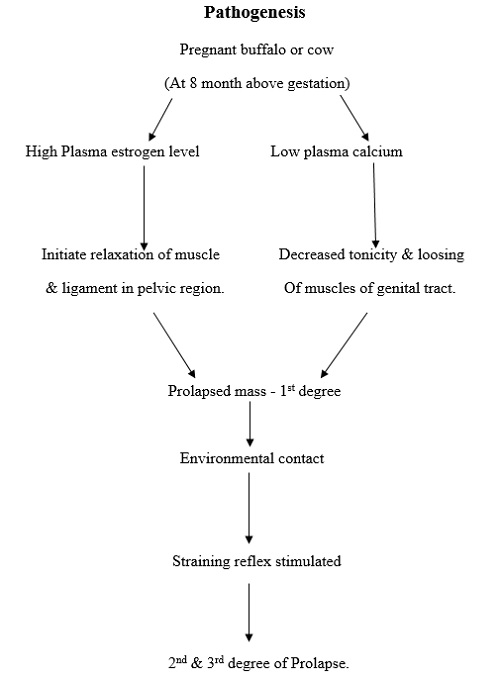
The incidence may be up to 0.5-1.0 % of calving’s. It usually occurs immediate after calving however it may also occur several days after parturition. Uterine prolapse occurs when the previously gravid uterine horn becomes invaginated/ folded in after calving and protrudes from the vulva. It is also termed as eversion of uterus or casting of “wethers” or casting of calf bed. In layman term also called as Bhelly Nikalna or Phool Nikalna. The following predisposing factors have been suggested for uterine prolapse in the cows:
Etiology
- The following predisposing factors have been suggested for uterine prolapse in the cows.
- In cows most commonly the last two-three months of gestation large amount of estrogenic hormone is secreted by placenta as a result relaxation of the pelvic ligament and adjacent structure.
- A common cause of vaginal prolapse is the pressure and weight of a large uterus in late pregnancy.
- Invagination of the tip of the uterus, uterine atony, and lack of exercise has all been incriminated as contributory causes.
- Atony of reproductive tract.
- General weakness of an animal.
- Due to hereditary genetic factor.
- Due to injuries of birth passage at first/subsequent parturition.
- Over distention of abdomen.
- Excessive amount of loose pelvic fat.
- Due to twins & low calcium level.
- Prolonged dystocia.
- Fetal traction.
- Fetal oversize.
- Retained foetal membranes.
- Hypocalcaemia
Degree of Prolapse
Prolapse can divide in four degree for convenience of treatment.
- Mild degree (1st degree)
- Second degree (2nd degree)
- Third degree (3rd degree)
- Fourth degree (4th degree)
Mild degree (1st degree)
- Mucous membrane of ventral floor of vagina is just pushed out while animal is sitting and disappear on getting up.
- Edema and reddening is found. Cervix is not involved.
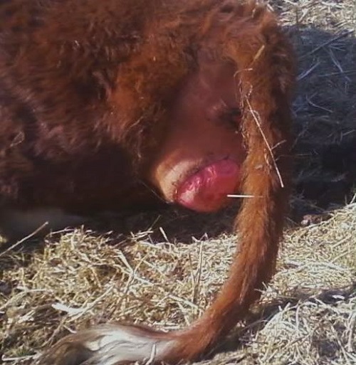
Second degree (2nd degree)
- Prolapsed mass remain persistently out and does not go back itself.
- Cervix is not involved. Slight more than 1st degree, edema found. Injuries might have occurred. Mucous membrane becomes necrotic in neglected case.
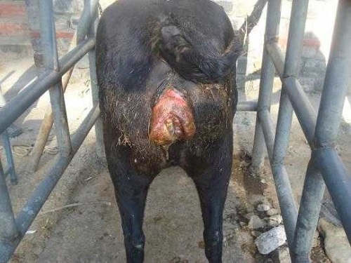
Third degree (3rd degree)
- Cervix already involved in prolapsed mass seen outside.
- Cervical seal is intact and seal can be seen. Necrosis, wound and edema of mucous membrane of vagina are noticeable.
- Blackening may found due to arrested venous blood flow suggestive of severe and long standing case.
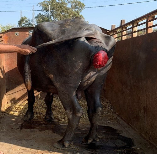
Forth degree (4th degree)
- Cervical relaxation occurs and cervical seal is lost and abortion/pre-parturition may occur within 24-72 hours.
- Necrosis of mucous membrane and tissue change are seen.
Principle for Management of Prolapse
- Checking increasing intra-abdominal and intra-pelvic pressure to avoid tenesumus and excessive repulsive force over the organ using sedative and analgesic.
- Prevention of chances of infection.
- Careful reposition of organ avoiding further injury.
- Management for retention of prolapse. Buhner’s technique and application of truss are practical technique.
- Prevention of reoccurrence of prolapse using medicaments. Viz. calcium therapy, progesterone therapy, use of anti biotic, and use of anti inflammatory as well as anti histaminic drugs etc.
- After care of the animals is of much importance to restore good fertility if the animal is to be maintained in the herd.
- Cow should be removed from the herd, and placed at platform raising rear quarters 2-6” inches higher than the fronts quarters.
Methods for Retention of replaced mass
- Vulvar truss
- Vulvar sutures:
- A burried/hidden purse string suture:
- Caslicks operation:
- Minchev’s method: (vaginopaxy)
- Cervical fixation:(cervicopaxy)
- Reefing operation:
- Buhner’s technique:
- Removal of large amount of perivagina fat
Treatment
- Administer low caudal epidural anesthesia using 5-10cc of 2% procaine/xylocaine hydrochloride solution to prevent tenesmus.
- Wash the prolapse organ with mild soap solution & water and rinse thoroughly with anti septic solution like PP solution.
- Empty the bladder by lifting up the prolapse mass or by catheterization.
- Reduce congestion by applying gentle pressure or by applying hypertonic salt solution.
- Apply antibiotic & emollients to prolapse mass & apply gentle pressure on prolapsed mass with palm over whole mass keeping in mind that further injury should not occur.
Prolapsed mass should be washed to make it free from dirt & debris with a mild, non irritating antiseptic solution or physiological saline. The specific treatment of uterine prolapsed is the subject of much debate and there is considerable individual variation about the techniques and approach of case. However, it must cover under than three major steps viz. Reduction of size, Replacement in the original position and Retention of prolapsed. A caudal epidural anesthetic is administered to relive tenesmus, allow easier reduction and replacement of the prolapsed mass and ensure painless procedure to animal.
Reduction, Replacement and Retention of prolapsed mass
The cow should be positioned in a manner to facilitate replacement of the prolapsed mass. As stated earlier, it is possible to replace the prolapsed in the standing animal. Animals that are recumbent should be placed in sterna recumbency, with their hind legs pulled behind them- the ‘frog-legged position’. Pulling the hind legs caudally provides a mechanical and gravitational advantage by tipping the pelvis forward. The placenta should be removed gently, if retained, as the edematous placentomes allow easy separation of the cotyledons from caruncles
Following removal of the placenta the uterine surface should be cleansed with dilute antiseptic solution. Fomentation with ice, hypertonic solutions of sugar or any water absorbent can be helpful in reducing the size of mass. The prolapsed organ should be elevated to the level of the ischium; this enables easier reduction and helps relieve vascular compromise and retention of urine. Elevation can be achieved by having one or two assistants suspend the organ in a sheet or towel, or by having an assistant sitting on the cow’s sacral region, facing backwards holding the organ upwards.
Replacement begins at the cervical pole of the organ. The organ is gently pushed back in to position, taking care not to traumatize the friable endometrium or uterine wall. Once replaced it is important to ensure complete eversion of the horns. Some practitioners will use a bottle as ‘arm extension’ to help ensure complete eversion of the horns. Following replacement, temporary closure of the vulva with a purse string or buhner’s sutures can be performed for retaining prolapsed mass. The tope truss can also be used for retention. Administration of oxytocin prior to replacement may facilitate the reduction.
Hysterectotomy/ Amputation of Prolapsed Uterus
It is advised only when replacement is impossible or when it is quite certain that replacement of a badly horn, lacerated, necrotic, infected uterus would result in death.
Post-operative care with the systemic injections of antibiotic, analgesics, anti-inflammatory, anti histaminic as well as local dressings at sutured site and general management is also very important after correction of prolapsed. Supplementation of utero-tonics like calcium supplements, oxytocin etc. may hasten the recovery. Animal should be housed in such floor so that hind portion will be elevated and the diet should be housed in such floor so that hind portion will be elevated and the diet should also restrict for few days. Animals should allow gently walking and avoid any cause of straining.
Prognosis
Prognosis is generally favorable for uncomplicated cases where there is no serious trauma or damage to the uterus encountered. Primiparas animals were found to have a better survival rate. The favorable prognosis found in the absence of severe hypocalcemia. Despite the favorable prognosis it is also important to consider the long-term effects on fertility. It may also increase the culling rate for infertility on subsequent fertility. The calving-to-conception interval may be longer in animals treated for uterine prolapsed.
| The content of the articles are accurate and true to the best of the author’s knowledge. It is not meant to substitute for diagnosis, prognosis, treatment, prescription, or formal and individualized advice from a veterinary medical professional. Animals exhibiting signs and symptoms of distress should be seen by a veterinarian immediately. |





Very good subject matter increaseing knowledge about prolapse