Abstract
A 7 yrs old Bullock was presented with protruded tongue, blood tinged excessive salivation having deep lacerated wound starting from the body to apex of tongue. The cause of injury might be licking of the iron chain of the other animal tied nearby as this bullock have vice to lick the ring of iron chain. The diagnosis was done by clinical examination. Pre surgical treatment was done to stabilize the animal and the traumatic portion of the tongue was surgically corrected under of sedation with Xylazine hydrochloride @ 0.1 mg/kg BW IM and local infiltration of 2% lignocaine hydrochloride. The mouth gag was applied and sutured with absorbable synthetic suture material. Simple interrupted suture techniques were used during surgery. Post-operatively, antibiotics and analgesic were given and advised to owner for offering soft feed. Animal recovered fast as healing of the oral cavity was very rapid.
Key words: Tongue, Tongue trauma, Tongue surgery, Deep Lacerated Tongue, Bullock.
Introduction
Most of the Vertebrates have tongue like Amphibians, reptile, birds and mammals. In mammals like as ruminants, dogs and cats, the tongue has various functions irrespective of that is also used to clean the fur and body by licking. The tongue is an important accessory organ in the gastro-intestitinal tract of animals and human being, as its helps in various important functions like in chewing the food against the hard palate, during mastication and manipulation of food for softening prior to prehension the food.
A ruminant uses its tongue to harvest forages during grazing or to consume harvested feed stuffs. Cattle consume forages during grazing by wrapping their tongue around the plants and then pulling to tear the forage for consumption. On average, cattle take from 25,000 to over 40,000 bites to harvest forage while grazing each day (Jane P., 2011, Feb).
The tongue is situated on the floor of the mouth, between the rami of the mandible. Tongue has three parts of a root, a body and the tip. The root is the caudal part and it is attached to the hyoid bone, soft palate and pharynx. Only its upper surface is free and slopes downwards and backwards.
The body has three free surfaces. The upper face is rounded and the lateral faces are flattened. The inferior face is related to the lingual muscles. Tip of the tongue is free and somewhat pointed. The dorsum linguae are the dorsal surface of the tongue. It is free throughout and is in contact with the palate except at the glosso-epiglottic space. The caudal part of the dorsum presents a remarkable eminence, the torus linguae, which is sharply defined in front by a transverse depression. The various structures presents as on the tongue
- Mucous membrane
- Muscles and glands
- Vessels and nerves
On the surface of the mucous membrane various kinds of papillae are
- Filiform papillae
- Fungiform papillae
- Circumvallate papillae
- Vallate papilla
The fungiform and vallate papillae are furnished with taste buds .The lingual glands are present in the sub mucous tissue. The lingual muscles are well develop (VAN)

Case History
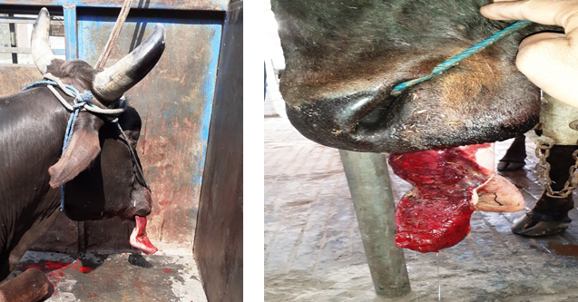
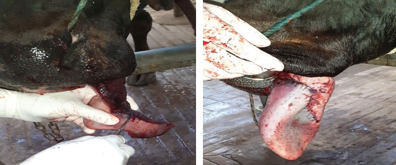
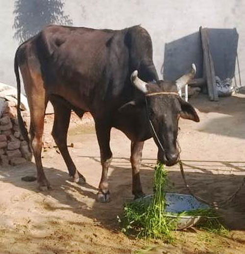
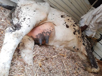
A 7 years old cow bullock was presented with a lacerated protruded tongue and had history vice of licking iron chain usually which was used to tie the other animals nearby. A day before presenting the case, the bullock had trauma on his tongue with the iron chain by wrapping their tongue around the ring of that iron chain. The owner saw the bleeding tongue of bullock. Clinical examination of oral cavity, revels transverse fine laceration of the dorsal and ventral portion which start from the body to apex of the tongue .It involving about 75% breadth of the tongue along with clinical signs of copious haemorrhagic ptyalism, blood tinched salivation, protrusion of tongue from centre of mouth (Fig1, 2 and 3.), inability to consume feed and drink water since trauma occur, dull and depress. The physiological parameters like Heart Rate, Respiration Rate and Mucous Membrane found in normal range
With rise in rectal temperature and mild dehydration seen. As bleeding wound margins indicated the viability of the tongue, it was decided for surgical repair (Dwivedi et al, 2013).
Surgical Treatment
The bullock was prepared for aseptic lingual surgery .Sedation was achieved with administration of Xylazine Hydrochloride @ 0.1 mg/kg and Inj. Atropine Sulphate @0.04 mg/kg BW I/V. The animal was restrained in cattle crush in standing position. After applying the mouth gag, Local anaesthetic 2% Lignocaine HCL was locally infiltrated at the margins of trauma and, the oral cavity is lavaged with diluted povidone iodine solution. The wound margins of lacerated tongues were surgically debrided of necrotic, blood clots and contaminated tissues (Fig-4).
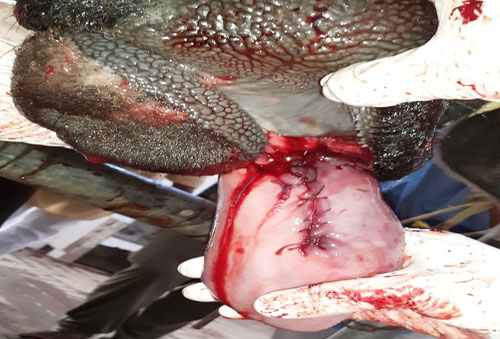
Suturing was performed using synthetic suture material simple interrupted pattern on both dorsal and ventral surface of tongue separately (Fig:-5&6). The sutures ensured obliteration of the dead space and proper apposition of the lingual mucosa on the dorsal and ventral surfaces (Jena.B et al,2017).
Post-operative
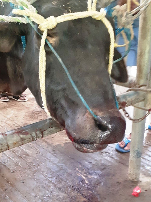
After completeion of the surgical procedure,the bucllock was administrated Inj.N.S.S. 7 L per day and Inj.DNS 10% 2L per day for 3 days to overcome the dehydration, Inj. Meloxicam @ 0.5mg/kg B.Wt, for 5 days, Inj. Enrofloxacin @ 10mg/kg B.Wt For 7 days, Inj. Antihistamine @0.2 mg/kg BW I/M regularly for 5 days. The owner was advised to lavaged the oral cavity with diluted Pot. Permagnate solution twice daily. The animals was offered Gur (Jaggery) and wheat dailya (porridge) daily and avoid feeding wheat straw for 10-12 days. To keep the rumen activity normal, Bolus. Gut-Boost(@ a product of Zenegal Neutraceuticals ) @2 BID for 7-10 days with jaguarry. After 12 days, the bullock was gradually shifting to green fodder, after 8 days later the owner reported normal prehension by the bullock. (Fig:-6)
Discussion
Lacerations, wounds and other lesion, may be traumatic or disease related normally heals without any surgical intervention by using daily mouth lavage and systemic antibiotics and by feeding a soft diet, however in severe traumatic cases that involve body of the tongue require surgery. Surgical intervention is indicated to promote healing and prevent deformity, to amputate a severely compromised apex, and to alter a scar or defect that is unacceptable to the owner (Jena.B et al,2017). As its well known, the tongue crucial role in grazing, chewing and in prehension of food, as much of the tongue as possible should be preserved. Primary closure of tongue lacerations in cows should be encouraged and partial glossectomy should only be the last resort (Dixon and Gerard, 2006; Duchrame. 2004; Dwivedi et al., 2013; Patel et al., 2013).
Conclusion
Blood tinched salivation during tongue trauma or injury is common because of interference with swallowing of saliva due to inflamination of the associated parts of the buccal cavity due to injury As in this case the laceration is deep and was on the body of the tongue due good vascularity of tongue prompts the fast and smooth recovery. Deep laceration of the tongue was successfully managed surgically resulting in rapid healing of the tongue was observed due to good vascularity (Thyagi, et al. 1993)
Reference
- B. Jena, S. Sahoo, S. Mahalik, A. Padhan, S. Nath (2017).Surgical repair of traumatic laceration of tongue in a cow. Journal of Livestock Science (ISSN online 2277-6214) 8: 131-133
- Dixon, P. M., & Gerard, M. P. (2006). Oral cavity and salivary glands. In: Equine surgery. Auer, J. A., & Stick, J. A 3rd Eds. WB Saunders.Elsevier Inc..Pp: 338-339
- Dwivedi, D. K., Gupta, A. K., Kushwaha, R. B., Gupta, P., & Sharma, A. (2013). Surgical repair of traumatic laceration of tongue in a pony. Indian Journal of Veterinary Surgery, 34(2), 148-148.
- Duchrame, N.G. (2004) Surgery of the bovine digestive system. In: farm animal surgery, Fubini, S. L. and Duchrane N.G. (eds) 1st ed, Elsevier (USA) St. Louis, Missouri, Pp: 164 – 165.
- JaneParish, (2011,Feb) Beef Production strategies Retrieved from https://extension.msstate.edu
- Patel, J. B., Patel, P. B., & Avasthi, H. A. (2013). Surgical Management of Tongue trauma in Mare. Intas Polivet, 14(1), 167-168
- Thyagi RPS, Jitsingh. Text book of Ruminant surgery, Hissar. 1983, 18.
- Van-Veterinary Splanchnology and Applied Anatomy Retrieved from http://ecoursesonline.iasri.res.in
|
The content of the articles are accurate and true to the best of the author’s knowledge. It is not meant to substitute for diagnosis, prognosis, treatment, prescription, or formal and individualized advice from a veterinary medical professional. Animals exhibiting signs and symptoms of distress should be seen by a veterinarian immediately. |


nice presentation
Nice presentation
Nice presentation, nice work and informative and good work..
very informative article.very nice presentation
Very informative article.