Abstract
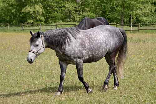
Leukoderma or Vitiligo is an autoimmune disease of the skin that is characterized by patchy depigmentation (i.e., white spots) and results from the loss of melanocytes, which are pigment-producing cells. Leukoderma is patchy depigmentation of skin with premature graying of local hairs with the usual manifestation as appearance of patches of gray or white hair. Leukoderma as a pigmentary disorder of unknown cause characterized by depigmented patches due to destruction of melanocyte. Causative factors of leukoderma may include environmental insult, autoimmune response in which melanocytes are destroyed by immune system and inherited predisposition and/ or copper deficiency. Treatment with injection Minshot, combined injections of copper, zinc, selenium and manganese, showed promising results and can be used in field to treat leucoderma in animals.
Keywords: Leukoderma (Vitiligo), Depigmentation, Causative Factors, Treatment.
Introduction
Leukoderma is defined as an acquired, chronic, depigmentation disorder characterized by white patches ‘snowflakes’. Leukoderma means loss of normal skin pigmentation (Radostits et al., 2007). It may be congential or acquired. Congenital or hereditary leukoderma develops due to lack of melanocytes or failure of melanocytes to produce melanin or failure of transfer of melanin to epidermal cells. It can also be acquired through loss of existing melanin or melanocytes (depigmentation). The exact mechanism of depigmentation of skin includes breakdown in the conversion of tyrosine to melanin because of reduced tyrosinase activity resulting in achromotrichia and leukoderma in copper deficiency (Underwood, 1977).
Historical Perspective
The first case series of leukoderma in dogs was reported in 1971. The first published case reports of feline leukoderma were reported in 1986. The most detailed report on feline leukoderma (in a Siamese Cat) was published in 1994. In the Horse, Duerst was cited to have first mentioned the term leukoderma in 1931.
Pathogenesis
The pathogenesis of leukoderma is multifactorial, and includes three main factors: genetic (Panduranga Rao et al., 2002), immunological (Fishman et al., 1997), and environmental (Cumming & Nordlund, 1995). Clinically, environmental factors are important in the development of vitiligo. Trauma, eczema, chemical agents, and fragility of keratinocytes play a role in development of vitiligo, so treatment decisions should be made taking these factors into account (Alikhan et al, 2011; Lee et al, 2005).
Types of Leukoderma: Leukoderma can be:
- Generalized, which is the most common type, when macules appear in various places on the body.
- Segmental, which is restricted to one side of the body or one area, such as the legs or face.
- Mucosal, which affects mucous membranes of the mouth and/or the genitals.
- Focal, which is a rare type in which the macules are in a small area and do not spread in a certain pattern within one to two years.
- Trichrome, which means that there is a white or colorless center, then an area of lighter pigmentation, and then an area of normally coloured skin.
- Universal, another rare type of vitiligo, and one in which more than 80% of the skin of the body lacks pigment.
Clinical Findings: Leukoderma signs include:
- Patchy loss of skin color, which usually first appears on the legs, face, and areas around openings and the genitals.
- Premature whitening or graying of the hair on the scalp, eyelashes.
- Loss of color in the tissues that line the inside of the mouth and nose (mucous membranes).
Depending on the type of leukoderma, it may affect
- Nearly all skin surfaces. With this type, called universal vitiligo, the discoloration affects nearly all skin surfaces.
- Many parts of the body. With this most common type, called generalized vitiligo, the discolored patches often progress similarly on corresponding body parts (symmetrically).
- Only one side or part of the body. This type, called segmental vitiligo, tends to occur at a younger age group.
- Only or only a few areas of the body. This type is called localized (focal) vitiligo.
- The face and legs. With this type, called acrofacial vitiligo, the affected skin is on the face and legs, and around body openings, such as eyes, nose and ears.
Diagnosis: On the basis of:
- Anamnesis (History Taking).
- Physical Examination, looking carefully at the affected skin.
- Blood Tests mainly Haemoglobin (Hb), Packed Cell Volume (PCV), Total Leucocyte Count (TLC), Total Erythrocyte Count (TEC), Mean Corpuscular Volume (MCV), Mean Corpuscular Hemoglobin (MCH), and Mean Corpuscular Hemoglobin Concentration (MCHC). The serum sample was used for analysis of micro minerals viz. Copper (Cu), Iron (Fe), Zinc (Zn) and Manganese (Mn).
- Skin Biopsy of the affected skin to be examined.
Treatment
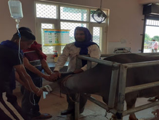
- Before the start of therapy deworming of all animal was done with Bolus Fenbendazole @5-7.5 mg/kg body weight. After 10 days of deworming observation pertaining to change in skin colour, hair colour, milk yield and fertility status were recorded at 10 days interval during course of treatment.
- Injection Intamin @ 10ml (total dose) subcutaneously at weekly interval.
- Treatment with injection Minshot, combined injections of copper, zinc, selenium and manganese, showed promising results and can be used in field to treat leucoderma in animals’.
Outcome
It is not possible to predict how an animal will respond to treatment. It is important to keep in mind that no one treatment works for every animal. Results can vary from one part of the body to another. Combining two or more treatments often give the best results.
References
- Alikhan, A.; Felsten, LM.; Daly, M. & Petronic-Rosic, V. (2011). Vitiligo: A comprehensive overview Part I. Introduction, epidemiology, quality of life, diagnosis, differential diagnosis, associations, histopathology, etiology, and work-up. J Am Acad , Vol. 65, No. 3, (September 2011), pp 473-491.
- Cummings, M.P. and Nordlund, J.J. (1995). Chemical leukoderma; fact of fancy. American Journal of Contact Dermatitis., 6: 122-127.
- Fishman, P.; Merimski, O.; Bharav, E. and Shoenfeld, Y. (1997). Autoantibodies to Tyrosinase; the bridge between melanoma nad vitiligo. American Cancer Society., 79: 126-130.
- Lee, AY,; Kim, NH.; Choi, WI. & Youm, YH. (2005). Less keratinocyte-derived factors related to more keratinocyte apotosis in depigmented than normally pigmented suction-blistered epidermis may cause passive melanocyte death in vitiligo. J Invest Dermatol., 124, No. 05, (May 2005), pp 976-983.
- Pandurangrao Rao, P.; Rama Dev.; Ravikumar, P.; Khan, P.M. and Kavitha, K. (2002). Suspected copper deficiency in buffaloes in Andhra Pradesh. Indian Vet J., 79: 1068-1069.
- Radiostits, O.M.; Gay, C.C.; Jincheliff K.W. and Constable P.D. (2007). Veterinary Medicine, 10th Sounders Elsevier Edinburgh, London.
- Underwood, E.J. (1977). Trace Elements in Human and Animal Nutrition, 4th Academic Press, London, pp 545.
|
The content of the articles are accurate and true to the best of the author’s knowledge. It is not meant to substitute for diagnosis, prognosis, treatment, prescription, or formal and individualized advice from a veterinary medical professional. Animals exhibiting signs and symptoms of distress should be seen by a veterinarian immediately. |


Is Vitiligo disease quarabull disease?
Very nice n informative Article