Abstract
The bovines are at a high risk of dropsical condition of fetal membranes mostly around the last trimester of gestation. The present review addresses dropsy of the fetal membranes that includes hydrops of amnion, hydrops of allantois and edema of chorio-allantois. The exact causes for dropsical condition was uncertain and require further research how the dropsical condition was developed in utero in gestation period. Various existing factor affecting the uterus, placental membranes and fetus responsible for formation of dropsical condition of fetal membranes, however some of these have been justified by logical interpretations. Considerable correlation exists between the fetal anomalies and degree of dropsy formation. This condition causes loss to the farmers in the form of fetal death, abnormal fetus and moreover gives poor prognosis to dam‘s life as well as fertility. Early diagnosis and addressing of treatment may give good prognosis without any complication.
Introduction
Gestational accidents are reported in all the species of animals and humans. Among the gestational accident dropsy of the fetal membranes is occasional occurrence, followed by affection of fetus, placenta and finally leads to danger to the dam. The pathological condition affecting the fetal membranes were hydramnios, hydrallantois and edema of chorio- allantois. As far as bovine species concerned, hydrallantois of fetal membrane is of great importance due to its incidence in field level, but in human hydramnios are most frequently encountered. Hydrops of the fetal membrane is the excessive level of fluid accumulation within the pregnant uterine horn, these pathological condition found singly but, rarely associated with each other in same animal. In uniparous animal the condition characterized by typically enlargement of abdomen, it may confused with hydroperitoneum (Ascites) and hydrometra in large and small ruminants respectively. If the condition not treated early in the advanced stage of pregnancy may result in animal unable to rise and gives poor prognosis to the dam‘s life.
Hydrallantois
In bovines allantois formed at second and third week of gestation period, however allantois is fully formed in large domestic animals by 24 to 28 days after conception. Fluids that filled in the allantoic cavity called allantoic fluid, that cavity stores the waste product of fetus which passes from fetus bladder through urachus. Amount of allantoic fluid varied from species to species that were tabulated.
|
Species |
Volume of Allantoic fluid at end of gestation |
| Cow |
9.5lit |
| Sheep and goat |
0.5-1.5lit |
| Dog |
10-50ml |
| Sow |
100-200ml |
| Cat |
3-15ml |
Hydrallantois is the gestational pathological condition of the fetal membrane characterized by rapid excessive accumulation of watery, amber colored fluid filled in the allantoic cavity over a period of 5 to 20 days in late gestation. Hydrallantois encountered about 85-90% of the dropsical conditions affecting the fetus and fetal membranes
Incidence
That records explains hydrallantois affects both heifer and parity animals of all species with irrespective of breeds. But, reports on hydrallantois in swine species was scanty.
Etiology: Etiology of the condition was divided into 3 groups.
| Primary causes |
|
| Secondary causes |
|
| Miscellaneous causes |
|
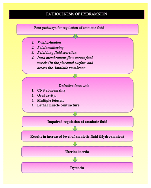
Hydramnios
Amnion formed about 13 to 16 days after conception in ruminants and equine, slightly earlier in pet animals (Dog, cat) and pig. It is a double walled sac that surround the fetus except at the level of umbilical ring. Amniotic fluid volume varies from species to species. Amniotic fluid mucoid in nature which get from saliva and naso-pharyngeal secretion of fetus. Volume can be regulated by fetal urination, fetal swallowing and fetal lung fluid secretion, intra membranous flow across fetal vessels on the placental surface and across amniotic membrane.
| Species |
Volume of amniotic fluid at end of gestation. |
| Cow |
5-6 lit |
| Mare |
3-7 lit |
| Goat |
0.4-1.2 lit |
| Ewe |
0.35-0.70 lit |
| Sow |
0.04-0.20 lit |
| Dog, cat |
8-30 ml |
Incidence:
Hydramnion condition develops slowly from several days to over month, especially the last 6 weeks of gestation in cattle, occasionally in sheep and goat, rarely reported in pig, dog and cat. Very rarely reported in horse. The degree of the condition depend on the anomalies of fetus.
Etiology
The most common for this condition was fetal anomalies, central nervous system defect, diabetes mellitus , severe foetal anemia with high cardiac output, idiopathic hydramnion, twin gestation also support the hydramnion .
Clinical sign, Diagnosis, Differential diagnosis, prognosis of the both Hydroamnion and Hydroallantois.
| Components | Hydroallantois | Hydroamnion |
| 1. Incidence | About 85-90% of cases | About 5-10% of cases |
| 2. Abdominal enlargement | Develops within 5-20 days | Develops slowly over months |
| 3. Abdominal wall | Round with heavy distension and tense | Pear shape and less tense |
| 4. Per -rectal examination | Distended uterine horn palpated. But not able to palpate the foetus and placentomes. | Uterine horn difficult to palpate. But able to able to palpate the foetus and placentomes. |
| 5. Foetal characteristics | Fetus are normal occasionally defect in kidney and liver of foetus on post mortem. | Defect and anomalous fetus mostly found. |
| 6. Placental and caruncles characteristics | Distended, leathery, gelatinous, and hypertrophied placentomes. Cauliflower like caruncles. | Normal characteristics of placentomes, placenta , chorio- allantois. |
| 7. Refilling capacity | Rapid refilling capacity after allanto-centesis. | Slow or not refill again |
| 8. Differential diagnosis | Intestinal obstruction, ascites, rupture of bladder, tumour. | Ruminal tympany, Vagal indigestion, Right side displacement of abomasum. |
| 9. Complications | Abdominal hernia, dislocation of hip (Bloated bull frog appearance), Retention of foetal membrane, Metritis | Rarely complication observed. Foetal anomalous monster cause dystocia. |
| 10. Prognosis | Dam future fertility and life-guarded to poor. | Dam future fertility and life-fair to good. |
III. Edema of the Allantois-Chorion
It is characterized by severe edema of the allantois- chorion with thickness of 4 -6 inches, mostly associated with brucellosis in bovine species (Roberts, 2004), however it is rarely associated with hydroallantois condition.
Haemato-biochemical and electrolyte changes in dropsical conditions:
- Blood cellular components : Hydrallantois affected animals suffer from Normocytic normochromic anemia ,i.e. decreased Hb , PCV , TLC and RBC levels due to accumulation of metabolic waste product present in the allantoic fluid and post -operative uterine adhesions followed by internal hemorrhage.
- Liver profile: Normal parturition without complication in bovines has negligible influence on the serum plasma enzymes, however, following hydrallantois and its correction by medical termination of pregnancy or surgical correction, the activities of serum glutamate oxaloacetic transaminase (SGOT) and serum glutamate pyruvate transaminase (SGPT) were increased which might be due to uterine adhesions
- Renal profile : Enlarged abdomen causes stress to the animal leads to stress- induced decrease blood flow to the renal system may complicated into dehydration , shock , and renal changes or toxic substance produced from the dead fetus culminate renal insufficiency. Moreover renal specific creatinine levels are elevated.
- Cortisol level: Cortisol levels are increased in delayed cases with advanced respiratory complication in heavily distended abdomen.
- Blood glucose level: High maternal blood glucose level in dropsical condition associated with diabetic pregnancy in women.
- Electrolyte concentration: Maternal hyponatreamia in dropsical condition associated with diabetic pregnancy, however Ducommun, (1967) documented no changes in the calcium and sodium level at the same time Phosphorus levels were increased and magnesium levels were decreased.
- Fetal fluid electrolyte concentration: No recent reports regarding foetal fluid electrolyte concentration during dropsical condition, only in 1967 Docoummun recorded that calcium values were normal level whereas phosphorus levels were high. Sodium levels are not static in hydrallantois cases due to changes in the permeability of placental membranes.
Treatment:
Amnio-reduction method
Human in late gestation with large volume of amniotic fluid associated with respiratory complication may necessitate the amnio-reduction technique by using sterile intravenous tubing connected to the evacuated container. This procedure needed weekly (or) semiweekly in late gestation. However possibility of risk of membrane rupture, placental damage, preterm labor. Advanced technical knowledge needed to perform this procedure. So, this procedure not validated in veterinary practice especially large animals in field level.
Trans- cervical Foley’s catheterization
Trans-cervical foley‘s catheterization is equal to allantocentesis technique. In this method 18” Rusch catheter fixed at the internal os of the cervix with help of inflating balloon attached with it, followed by administration of prostaglandins analogue and dexamethasone leads to slow drainage of allantoic fluid occur after 48 hours of treatment. If improper fixing (or) complete damage to the allantoic cavity leads to sudden release of allantoic fluid may end in hypovolemic shock. Hypovolemic shock can be managed by administration of intravenous hypertonic saline solution.
Cervical negotiation and oxytocin administration
Trans-cervical Foley’s catheterization alone not give complete response to hydrallantois associated with uterine atony, for combat the uterine atony non-violent contraction of uterus were achieved with administration of 20 IU of oxytocin four times at 20 minutes interval results in drainage of allantoic fluid slowly.
Abdomino-centesis
This method is not a complete choice for termination of hydrallantois in large animal but some extend possible in sheep and goat for relieving respiratory distress, 16 gauge needle, trocar and canula used for relieving the allantoic fluid, followed by induction with luteolytic drug.
Medical termination of pregnancy
Termination of pregnancy was the best choice of treatment when compared to other method, moreover primarily MTP was combined in all technique of treatment method. Elsewhere scientist mostly tried with combination of prostaglandins and Dexamethasone combination of pregnancy as compared to single drug alone. Prostaglandins groups like Dinoprost (25mg-total dose I/M), cloprostenol (500µg- total dose I/M). Either one of the prostaglandins combined with Dexamethasone (50-60mg). The termination of pregnancy attributed by administration of dexamethasone encourage fetal corticosteroids and stimulate 17-alpha hydroxylase enzyme in the placenta and prostaglandins causes luteolysis of functional corpus luteum.
Cesarean section
Medical termination of pregnancy not always gives complete results due to failure of ripening of cervix and incomplete cervical dilation, it necessitate the cesarean section to solve the condition. Precautionary measure during cesarean section is slow drainage of allantoic fluid by using sterile rumen trocar and simultaneous administration of hypertonic solution intravenously to combat the hypovolemic shock.
Conclusion
Dropsical condition is one among the major impediment hindering the future fertility and life of the dam. It is well known problem in the field level for veterinarian to manage the condition, because of life threatening hypovolemic shock when sudden drainage of the fetal fluid. Various unknown causes are interplay for the formation this condition in late gestational period. Early diagnosis of this condition in the field level through per-rectal and per-vaginal examination in concordant with history of pregnancy positive at first or second trimester pregnancy could supportive to the differentiation other gestational accident. According to the clinical status of the animal the treatment protocols were modulated from medical termination pregnancy alone or combination with catheterization technique to solve the condition. The hopeless delayed cases with respiratory complication and lateral recumbency the prognosis of the animal was guarded. Post-obstetrical treatment for this condition might have to be administration of antibiotics to combat the infections. Ecbolic drug, Calcium borogluconate to restore the uterine motility for expulsion of fetal membranes and its contents. Salvage the animal, if the condition suspected to be hereditary and discourage the rebreeding of this animal.
|
The content of the articles are accurate and true to the best of the author’s knowledge. It is not meant to substitute for diagnosis, prognosis, treatment, prescription, or formal and individualized advice from a veterinary medical professional. Animals exhibiting signs and symptoms of distress should be seen by a veterinarian immediately. |


I’m not that much of a online reader to be honest but
your sites really nice, keep it up! I’ll go ahead and bookmark your site to come back in the future.
Many thanks