Introduction
Coccidiosis is one of the economically important and major parasitic diseases in the calves caused by Eimeria spp. which are often underemphasized in Indian scenario. It is responsible for considerable number of morbidity and mortality particularly in calves aged upto 1 year. Majority of cattle are infected with coccidia, but only a limited number of them suffers from clinical coccidiosis (Khan et al., 2013). Coccidiosis is seen in late summer and winter months but it may occur throughout the year in India. Clinical coccidiosis is commonly prevalent in animals under the age of 1 year and susceptibility to infection gradually declines with advancement of age because of previous exposure and development of immunity (Nambiar and Devada, 2002). A negative correlation exists between the age of cattle and risk of infection and younger animals depicts the higher prevalence (27.23 %) of coccidial infection than older animals (15.65 %) (Cicek et al., 2007).
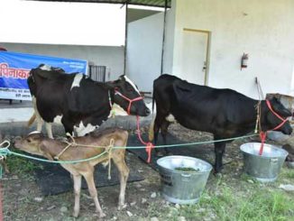
The disease is particularly a problem of confined animals kept under intensive husbandry practices and is more common in housed animals than in that practice semi-intensive. Overcrowding and lack of hygienic measures increases the chance of infection (Swarnakar et al., 2015). The major damage is due to the rapid multiplication of the parasite in the intestinal wall and the subsequent rupture of the intestinal cells. Several stages of multiplication occur before the final stage and the unsporulated oocyst are passed out in the faeces. These oocysts are extremely resistant to environmental stress and difficult to remove from the environment completely.
Etiology
Coccidiosis is an important intestinal disease of cattle caused by various Eimeria spp. There are more than 12 species of Eimeria and one species of Isospora have been described to infect cattle. However only three (E zuernii and E bovis) are most often associated with clinical disease under the field conditions while other species have been shown to be mildly or moderately pathogenic (Verma et al., 2018). Eimeria spp. parasitizes the epithelium of the digestive tract and the infection certainly damages the lining of the gut causing diarrhea and dysentery.
Transmission
The disease is transmitted by ingestion of sporulated oocysts mainly through faecal-oral route. Infected fecal material also contaminates feed, water or soil; therefore, cattle can acquire the disease by eating and drinking from contaminated sources or by licking itself or other animals. Calves may become infected by nursing contaminated udders (Reddy et al., 2015). Nevertheless, the severity of clinical disease depends on the level of number of oocysts ingested and the immune status of animal. The more oocysts ingested, the more severity of the disease expected.
Clinical Signs
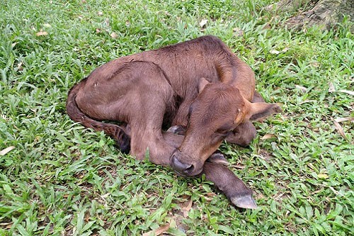
Coccidiosis in cattle commonly occurs as subclinical disease and causes substantial economic losses. The signs of clinical coccidiosis are reduced appetite, decreased body weight, unthriftness, rough hair coat, dehydration, weakness, dry muzzle, pale congested conjunctival mucous membrane, low body temperature, diarrhea, dysentery and blood mixed faeces attached to the perineum regions. The tenesmus and prolapsed rectal mucosa can also be observed in some cases (Radostits, 1994).
Diagnosis
- Clinical Signs: The signs usually occur about 17 days after the ingestion of oocysts and by the time clinical signs occur, the damage is far advanced. The history of cases presented frequently includes a preceding stressful event in the animal’s life.
- Faecal examination: Qualitative analysis of coccidiosis can be done by fecal examination by flotation or smear which shows the presence of large number of oocysts. For quantitative diagnosis McMaster technique is adopted (Zajac and Conboy, 2006).
- Postmortem examination: Pinpoint haemorrhages in the wall of intestine can be noticed. Intestinal scrapping has to be observed under microscope for the presence of schizonts, merozoites or gametes.
Differential diagnoses
It includes bovine viral diarrhea, salmonellosis, malnutrition, toxicity, or other intestinal parasites (Verma et al., 2018).
Treatment
Affected calves are isolated to prevent further contamination of the premises and are treated with 33.33 % (w/v) Sulphadimidine @ 100 mg/kg body weight IV for 7 days (Reddy et al., 2015) or Sulphadimidine @ 200mg/kg body weight for 5 days or Amprolium at 10 mg/kg/day for five days. Supportive treatments such as fluid therapy, vitamins, antibiotics, anti-inflammatory and probiotics drugs are used to prevent secondary infections. Corticosteroids are contraindicated in coccidiosis because they increase the shedding of oocysts and induce clinical disease in sub-clinically infected calves.
Anticoccidial and Coccidiostats drugs for use in cattle (Richards et al., 2015)
|
Sl. No. |
Drug | Therapeutic |
Preventive |
| 1. | Amprolium | 10 mg/kg/day for five days | 5 mg/kg/day for 28 days |
| 2. | Sulfonamides | 200 mg/kg/day for five days | |
| 3. | Monensin | 1 mg/kg/day, 10-30 g/ton | |
| 4. | Lasalocid | 1 mg/kg/day, 10-30 g/ton | |
| 5. | Decoquinate | 0.5 mg/kg/day for 28 days or longer |
Economics of Coccidiosis
Coccidiosis is responsible for undesirable economic losses to the farm income in terms of mortality and morbidity particulalry in young calves (Nalbantoglu et al., 2008). The losses caused by coccidiosis can be divided into quantifiable and unquantifiable because the losses due to subclinical infections in calves probably far outweigh those of clinical disease. The quantifiable losses are from clinical coccidiosis i.e., impaired production, mortality ranging upto 24% and cost of treatment (Rodriguez et al., 2017). The unquantifiable losses are from subclinical infection i.e., impaired alimentary function, reduced feed conversion efficiency, reduced growth and significant increase in the susceptibility to other diseases because of poor immunity.
Prevention and Control
- Water and feed troughs should be kept clean and emptied on a regular basis
- The floors and holding areas should be disinfected regularly
- All bedding materials must be kept clean and dry
- Avoid overcrowding of calves and provide adequate ventilation
- Mixing of different age groups of animals particularly older cattle has to be avoided since they are big risk factor for spreading infection to younger calves
- Use anticoccidial drugs or coccidiostats regularly following prescribed recommendations
- Mass medication of the feed and water supplies to prevent new cases and to minimize the effects of an epidemic.
Conclusions
Coccidiosis is an economically important parasitic disease of calves reared in intensive animal husbandry conditions. It causes both quantifiable and unquantifiable losses to the farm growth. Hence treatment and control must be a two-fold approach: good animal husbandry practices to prevent the ingestion of oocysts and as well as the use of anticoccidial drugs to prevent the further disease and premises contamination.
References
- Bohrmann R. Treatment with toltrazuril in a natural outbreak of coccidiosis in calves. DTW. Deutsche Tierarztliche Wochenschrift. 1991; 98(9):343-5.
- Cicek H, Sevimli F, Kozan E, Kose M, Eser M, Dogan N. Prevalence of coccidia in beef cattle in western Turkey. Parasitol Res. 2007;101:1239–1243.
- Khan, M.N., Sajid, M.S., Abbas, R.Z., Zaman, M.A., Sikandar, A. and Riaz, M., 2013. Determinants influencing prevalence of coccidiosis in Pakistani buffaloes. Pakistan Veterinary Journal, 33(3).
- Nalbantoglu, S., Sari, B., Cicek, H. and Karaer, Z., 2008. Prevalence of coccidian species in the water buffalo (Bubalus bubalis) in tRakshithhe Province of Afyon, Turkey. Acta Veterinaria Brno, 77(1), pp.111-116.
- Nambiar KS, Devada K. Prevalence of bovine coccidiosis in Trissur. Indian Vet Med J. 2002;26:211–214.
- Radostits OM, Blood DC, Gay CC. A Textbook of the Diseases of Cattle, Sheep, Pigs, Goats, and Horses. 8th ed. London. Philadelphia, PA: Bailliere Tindall, 1994, 1181-1199.
- Reddy, B.S., Sivajothi, S. and Rayulu, V.C., 2015. Clinical coccidiosis in adult cattle. Journal of parasitic diseases, 39(3), pp.557-559.
- Richards, C., Step, D.L. and Giedt, E.J., 2015. Coccidiosis treatment and prevention in cattle. Oklahoma Cooperative Extension Service VTMD-9129
- Rodríguez-Vivas, R.I., Grisi, L., de Leon, A.A.P., Villela, H.S., de Jesús Torres-Acosta, J.F., Sánchez, H.F., Salas, D.R., Cruz, R.R., Saldierna, F. and Carrasco, D.G., 2017. Potential economic impact assessment for cattle parasites in Mexico. Review. Revista Mexicana de Ciencias Pecuarias, 8(1), pp.61-74.
- Swarnakar, G., Bhardawaj, B., Sanger, B. and Roat, K., 2015. Prevalence of gastrointestinal parasites in cow and buffalo of Udaipur district, India. Int. J. Curr. Microbiol. App. Sci, 4(6), pp.897-902.
- Verma, R., Das, G., Saiyam, R. and Bendigeri, S., 2018. Clinical coccidiosis in calves and its treatment. Journal of Entomology and Zoology Studies. JEZS 2018; 6(2): 2964-2967
- Zajac MA, Conboy GA (2006) Faecal examination for the diagnosis of Parasitism. 7th edn. Blackwell Publishing, Ames, pp 3–10
| The content of the articles are accurate and true to the best of the author’s knowledge. It is not meant to substitute for diagnosis, prognosis, treatment, prescription, or formal and individualized advice from a veterinary medical professional. Animals exhibiting signs and symptoms of distress should be seen by a veterinarian immediately. |

Be the first to comment