Introduction
Theileriosis is considered as one of the important disease which causes devastating economic losses in livestock production. The disease is known as East coast fever, Tropical theileriosis and Oriental theileriosis caused by Theileria annulata and Theileria parva respectively transmitted by ixodid ticks (hard ticks). Theileria spp. involves a complex life cycles in both vertebrate and invertebrate host to form sporozoites. In white blood cells, the sporozoites develop into schizonts and the schizonts develop into merozoites Red blood cells. The geographic distribution of East coast fever is reported to occur in Sub-Saharan Africa and Theileria annulata occurs in Southern Europe and Middle East. The presence of Theileria orientalis is widespread. Theileriosis is characterized by anorexia, emaciated, fever, high body temperature, pale mucous membrane, generalized lymphadenopathy, nasal discharge, decreased milk production and diarrhoea. The most common post-mortem findings in the affected animals are ecchymosis and petechiae in the serosal surfaces of internal organs. Theileriosis is diagnosed on the basis of clinical symptoms and microscopic examination of blood smears. Therapeutic management of Theileriosis include antiparasitic drug like Buparvaquone @2.5mg/kg body weight. Since, Theileriosis affects adversely in the livestock population and production so proper management and control should be adopted to prevent such fatal and harmful protozoal infection in bovines.
Definition
Theileriosis is a tick borne protozoan infection caused by Theileria spp. in cattle, sheep, goat and horses which is characterized by fever, pale mucous membrane, general lymphadenopathy, dysopnea, decreased milk production and skin lesions.
Etiology
The disease Theileriosis is caused by the diverse species of theileria which belongs to phylum apicomplexa, family theileridae and genus theileria. East coast fever is the most common and economically important disease occurs in cattle caused by Theileria parva and transmitted by ticks Rhipicephalus appendiculatus. Tropical theileriosis, also known as Mediterranean coast fever is caused by Theileria annulata. In this case Hyalomma anatolicum act as main vector for spread of disease. Hyalomma anatolicum is a three host tick which transmit the disease transtadially. Haemophysalis spp. act as main vector for oriental theileriosis also known as Japanese theileriosis, which is caused by Theileria orientalis genotype ikeda.
Epidemiology
The occurrence of East coast fever is mainly in Eastern, Central and Southern Africa which affects both cattle and buffalo. The prevalence rate of East coast fever is highest in wet areas which is high favorable for the development of the ticks. There can be two cases of endemic scenario, either it can have high prevalence but low fatality or high fatality with low prevalence. The disease is eradicated from Southern Africa to Zambezi River and epidemics of East coast fever occur when there is failure in tick control. The geographic distribution of Theileria annulata is Southern Europe, Middle East and parts of Asia. The case fatality rate of Tropical Theilerosis is high in exotic cattles (20-90% mortality).
Modes of transmission
Theileriosis is a tick borne disease in which protozoa is transmitted transstadially via Rhipicephalus and Hyalomma spp. via ixodid ticks. East coast fever, Tropical theileriosis and Oriental theileriosis is transmitted by Rhipicephalus appendiculatus, Hyalomma anatolicum and Haemophysalis spp. respectively. Theileriosis does not involve transovarian transmission. The protozoa undergo its developmental stages in the vector (tick) and the tick transmit the parasite transstadially via larva, nymph and adult stages.
Pathogenesis
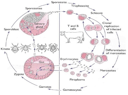
The completion for the life cycle of the parasite in the mammalian host, the theileria consecutively utilize both white blood cells and red blood cells. The sporozoite stage of the theileria transmitted through the salvia of infected ticks to the host. Sporozoites enter the leucocytes and they develop to schizonts within a few days. The most pathogenic spp. multiply in the host’s white blood cells and the less pathogenic spp. multiply in the host’s red blood cells. The schizont stage of pathogenic spp. divides the host’s white blood cells and as a result the parasite circulated throughout the lymphoid tissue. In due course of the disease, the schizonts multiplies and undergo merogony hence, releases merozoites which infects red blood cells and produces piroplasms. When the tick bites the infected host along with the red blood cells the piroplasm also enter in the gut of the tick and further these piroplasm undergo sporogony and forms sporozoites migrates to the salivary gland and the ticks transmit the infection transstadially to their next host.
Clinical findings
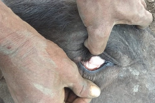
The primary clinical symptoms in East coast fever are enlargement of lymph nodes, increased body temperature (40-41ᶿC), diarrhoea. In later stages, nasal and ocular discharge, salivation, generalized lymph node enlargement, enlargement of spleen (splenomegaly), after 1-2 days there is anorexia, emaciation and decreased milk production in lactating animals. Brain involvement have been reported occasionally which is characterized by circling movement Therefore, East coast fever is also known as Turning sickness or cerebral theileriosis. Periodically, ocular diseases (corneal opacity and exopthalmia) and skin lesions (nodules, hemorrhagic and ulcerative lesions) also be seen.
The clinical findings in Tropical theileriosis are similar with East coast fever. The signs include anemia, pale mucous membrane, jaundice and hemoglobinuria, convulsions, torticollis, tachycardia, labored breathing. In severe cases, hemorrhagic diarrhoea is seen and sometimes animals may suffer from abortion in tropical theileriosis.
Clinical pathology
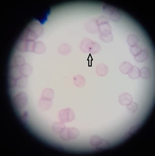
Examination of blood shows panleukopenia, thrombocytopenia and lymphocytopenia. The blood smears examination and lymph node biopsy will show schizonts in lymphocytes and piroplasms in erythrocytes. Anemia is the most striking features in Tropical theileriosis, along with haemoglobinuria, bilirubinuria and bilirubinemia is evident. Depletion in White blood cells and platelet counts is more severe in East coast fever than Tropical theileriosis. Oriental theileriosis in cattle causes marked reduction in red blood cell count, haemoglobin concentration, packed cell volume and increase in mean corpuscular value.
Necropsy findings
The most prominent lesions in East coast fever during the post- mortem of the animal are pulmonary edema, hyperemia and emphysema. Enlargement of lymph node, spleen and ulceration of abomasum and intestines is observed. Haemmorhages and necrosis are obvious in variety of tissues and organs like lungs, liver, kidney, GIT etc. The pseudo infarcts found to be present in the kidney, liver and alimentary tracts. The small lymphoid nodules are present in the brain in cerebral theileriosis.
In Tropical theileriosis, the necropsy findings are alike to those of East coast fever but includes multicentric malignant lymphoma in the lymph nodes due to lymphoid proliferation.
Diagnosis
Giemsa-stained thin blood smears or lymph node biopsies can be done to detect the theileriosis in live animals. On microscopic examination of the blood smears reveal the presence of Koch Blue Bodies and Intra erythrocyticpiroplasm. Necropsy findings useful in which the schizonts can be detected in smears from lungs, spleen, liver and lymph nodes. Various types of serological techniques is available which includes Immunofluorescent antibody test (IFAT), Compliment fixation test (CFT), Enzyme linked immunosorbent assay (ELISA) and Indirect haemagglutination test. IFAT is most commonly used serological test to detect theileriosis in animals. The most widely used technology to identify the parasite is Polymerase chain reaction. PCR test is more sensitive and specific to detect carriers and to detect infected ticks.
Treatment
The drug of choice for theileriosis is Buparvaquone which is administered at the rate of 2.5mg/kg BW deep intramuscular route preferably neck muscle with 2 doses 48 hours apart and Oxytetracycline can be given at the rate of 10-20mg/kg BW Intravenously mixed with normal saline solution (NSS) but oxytetracycline have moderate efficacy.
A higher success rate is shown by halofuginone lactate and parvaquone, which were recently introduced for treatment. Oral administration of two doses of halofuginone lactate is quite effective at 1.2mg/kg BW for acute syndrome. In most of the cases intramuscular administration of two doses of parvaquone at 48 hours apart with 10mg/kg BW is mostly effective. The only precautions to be taken is that correct dose should be admen otherwise animal may become carrier of disease. Other supportive therapy such as NSAID, iron injection and liver supportive should be given.
Prevention and control
Culling of infected animal for healthy herd should be done immediately to prevent the spread of disease. Restricted movement should be their of infected livestock. Exposure to the main vector can be minimized by different methods of tick control such as acaricides and rotational grazing in endemic areas. Animals must be tested for blood protozoa prior to blood transfusion. In some countries, vaccines are available for East coast fever and Tropical theileriosis. Vaccine available for Tropical theileriosis is attenuated live vaccine, however for East coast fever several strains of Theileria parva are administered in combination with an antibiotic, which is preferably a long term tetracycline. The outcome of this administration is inapparent or mild infection which is followed by good immunity leading to formation of carriers. Initiation of live vaccine in anon-endemic area is a point of concern and should be avoided.
Conclusion
Theileriosis is an important vector born disease leading to high morbidity and mortality. The severity of the disease depend upon the species of Theileria, strain and host factors i.e. immunity and concurrent illnesses. The common virulent organisms in cattle are Theileria annulata, which causes tropical theileriosis, and T.parva, which causes East Coast fever, in sheep and goats is usually caused by T.lestoquardi (formerly T.hirci), T.uilenbergior T.luwenshuni. and T.parvaaffects cattle and water buffalo. Common clinical signs are swelling of the draining lymphnode Fever 40–41°C nasal discharge, Lacrimation swelling of the eye lids and ear anemia, Jaundice, Anorexia, dyspnoea diarrhoeain occasional cases of brain involvement occur and are characterized by circling, known as ‘turning sickness’ or cerebraltheileriosis. Theleriosis can be diagnosed by history of presence of Ticks, clinical findings such as lymph node swelling, anaemia, icterus post-mortem findings (Punched necrotic ulcer in Abomasum). In blood smear examination some time KBB (Common in T.parva) T.parvaschizonts seen in Lymphoblast & T.annulataschizont in macrophages. Buparvaquone considered as drug of choice for theileriosis and other supportive therapy provided for treating the affected animal.Control of vector, avoiding nutritional stress and effective vaccination (Rakshavac-T vaccine @3ml,s/c, primary vaccination at 2 months age & revaccination after 1 year for cross bred and exotic cattle)helps in prevention of the disease.
Reference
- A text book of preventive veterinary medicine-by Amalendu Chakrabarti
- Bhattacharyulu, Y., Chaudhri, R.P. and Gill, B.S., 1975. Transstadial transmission of Theileriaannulata through common ixodid ticks infesting Indian cattle. Parasitology, 71(1), pp.1-7.
- Gill, B.S. and Bhattacharyulu, Y., 1977. Bovine theileriosis in India. Theileriosis. Ottawa, IDRC, p.8.
- Kakati, P., Sarmah, P.C., Ray, D., Bhattacharjee, K., Sharma, R.K., Barkalita, L.M., Sarma, D.K., Baishya, B.C., Borah, P. and Stanley, B., 2015. Emergence of oriental theileriosis in cattle and its transmission through Rhipicephalus (Boophilus) microplus in Assam, India. Veterinary world, 8(9), p.1099.
- Morrison, W.I., 2015. The aetiology, pathogenesis and control of theileriosis in domestic animals. Rev Sci Tech, 34(2), pp.599-611.
- Uilenberg, G., Perie, N.M., Spanjer, A.A.M. and Franssen, F.F.J., 1985. Theileriaorientalis, a cosmopolitan blood parasite of cattle: demonstration of the schizont stage. Research in veterinary science, 38(3), pp.352-360.
- Velusamy, R., Rani, N., Ponnudurai, G., Harikrishnan, T.J., Anna, T., Arunachalam, K., Senthilvel, K. and Anbarasi, P., 2014. Influence of season, age and breed on prevalence of haemoprotozoan diseases in cattle of Tamil Nadu, India. Vet. World, 7(8), pp.574-578.
- Veterinary Medicine A textbook of the diseases of cattle,horses, sheep, pigs and goats by O. M. RadostitsC.C.Gay,K. W. Hinchcliff, P. D. Constable.
| The content of the articles are accurate and true to the best of the author’s knowledge. It is not meant to substitute for diagnosis, prognosis, treatment, prescription, or formal and individualized advice from a veterinary medical professional. Animals exhibiting signs and symptoms of distress should be seen by a veterinarian immediately. |


Be the first to comment