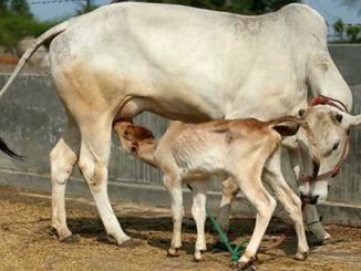
Post parturient Injuries and diseases in large animals
Obstetric trauma to the genital tract also predisposes to infection, and where severe contusion has occurred there is a marked risk of infection particularly by anaerobic bacteria. There are three important conditions that follow delivery of the fetus, namely prolapse of the uterus, retention of the afterbirth and puerperal infection, and are given special consideration elsewhere. In addition, there are numerous accidents and diseases that accompany or follow parturition. A difficult foaling may be followed by laminitis or tetanus, and in all species puerperal animals may incur embolic pneumonia, toxaemia, septicaemia and pyaemia as sequels to uterine infection. Endocarditis, unthriftiness and sterility are possible later sequelae.
Postpartum haemorrhage into the birth canal
Etiology
It is caused by accidental damage to the soft tissue walls of birth canal, especially the uterus and vagina. The damage may be caused by unguarded sharp fetal extremities or by excessive obstetric force. Significant haemorrhage from the capillary plexuses around the crypts can occur only when excessive force is used in early removal of cotyledonary type afterbirth. After removal of the fetus much blood may accumulate in the uterus before it begins to escape via the vagina; alternatively blood may drain through a tear in the uterine wall into the abdomen.
Clinical Signs
Mostly blood coming through vulva, after heavy traction or fetotomy followed by traction the anterior vagina and palpable parts of uterus are examined systematically by hand. Laceration of mucosa or palpation of a pulsating artery may indicate the site of haemorrhage.
Treatment
Visible and palpable bleeding points are subjected to pressure with clean towel or cotton wool along with alum. If a spurting artery is located it is clamped with artery forceps. An injection of oxytocin will cause uterine contraction and possibly some pressure on to the bleeding point. General symptoms of severe haemorrhage and shock can be counteracted by blood transfusion (4–5 litres) from a neighbouring animal.
Contusion and laceration of the birth canal and neighboring structures
Contusion of birth canal may occur during forcible extraction of the fetus. Cervix and vulva are more prone for injury than dilatable vagina, Infection with Fusiformis necrophorum and other pyogenc bacteria may lead to severe necrotic vaginitis and toxaemia.
Treatment
All vaginal contusions and lacerations should be treated with mild emollient and antibiotic preparations; parenteral antibiotics should also be given. Caudal epidural anaesthesia can be opted for temporary relief. Rupture of vagina and cervix must be repaired.
Haematoma of the vulva
unilateral or bilateral enlargement of vulval lips. Haematoma of the vulva may be confused with prolapse, tumour or cyst of the vagina. If left untreated, natural resolution usually occurs within a few weeks with resorption of fluid and regression of swelling; occasionally, pyogenic infection ensues and may be accompanied by fibrosis and distortion of the vulva, with vaginal aspiration. If left for 3 or 4 days after labour the haematoma may be safely incised and the clot removed without recurrence of haemorrhage. An abscess should be opened and drained.
Perineal injuries at parturition
This type of injury occur during the second stage of labour in both the cow and the mare, mostly in primiparous animals. These injuries may be classified as first-, second- and third-degree tears and rectovaginal fistulae. Tears which extend more deeply into the perineum do not close spontaneously and destroy the sphincteric effect of the vulva and lead to aspiration of air into the vagina i.e pneumovagina.
Damage to the lumbosacral plexus
When a large fetus is forcibly drawn into the maternal pelvis the lumbar nerves which pass over the lumbosacral joint to form the anterior part of the lumbosacral plexus may be damaged; paralysis of the gluteal or obturator nerves is a possible result. This is particularly likely when an oversized fetus becomes impacted in a state of ‘hiplock’, the nerves being trapped between the lumbosacral promontory of the mother and the iliac bones of the calf. In addition, the obturator nerve, as it passes down the inner surface of the iliac shaft, may be damaged by an oversized fetus.
Gluteal paralysis
Gluteal paralysis is seen in the mare and cow; in the mare it has followed spontaneous birth. It is recognised when the dam is found to have difficulty in rising and when she walks with ‘weakness of the hindlimbs’. Later, atrophy of the gluteal muscles is apparent. Prognosis is favourable, the disability usually disappearing in a few weeks, although occasionally complete recovery may take months. The animal may be helped to rise by lifting on its tail and then steadying its hindquarters. In order that the mare may suckle the foal and also rest on its feet, slings may be usefully employed. If the mare or cow cannot get up within a few days of parturition the prognosis is grave.
Obturator paralysis
Obturator paralysis is more frequent in cows than mares. The obturator nerve supplies the adductor muscles of the thigh; thus when both nerves are damaged the legs will be splayed and the cow is unable to rise. If the cow is helped to its feet, the legs slide out laterally. When paralysis is one-sided the cow also requires assistance to get up but can stand readily, if the affected leg is prevented from sliding outwards. If the cow falls there is a risk of limb fracture or dislocation of the hip joint. Where there is complete and bilateral paralysis, prognosis should be guarded; where it is unilateral and the animal can walk with assistance. Most cases show rapid improvement within a few days and progress to a complete recovery. Treatment comprises good nursing. The cow should be well bedded with short litter on an earthen floor or on a concrete floor on which sand or grit has first been sprinkled. She must be assisted and maintained on her feet for milking or suckling and as often as possible at other times. The patient should be stimulated to walk but should be prevented from falling awkwardly. Slings are occasionally employed for cattle. Bedsores must be prevented, hindquarters massaged, the bedding frequently changed and the cow’s rear and udder kept clean and dry.
Rupture of the Uterus or Vagina
Rupture of the uterus may occur spontaneously, but faulty obstetric technique is a more frequent cause. Spontaneous rupture is most likely to arise in association with uterine torsion or with cervical non-dilatation but is also possibly due to the gross uterine distension that occurs with twins in one horn, with hydrallantois or with excessive fetal size.
Breech presentation predisposes to rupture because the breech of the calf fully occupies the maternal pelvic inlet and allows no egress for the fetal fluid when the uterine and abdominal contractions build up the hydrostatic pressure within the uterus.
Prolapse of the Bladder
Prolapse of the bladder may follow a rupture in the floor of the vagina or eversion through the dilated urethra and may occur during or after parturition. The rounded organ protrudes from the vulva. The kink that forms in the urethra prevent micturition thus the organ progressively distends with urine.As treatment is concern the surface of the bladder is cleaned and the organ is punctured with a hypodermic needle to allow drainage of urine. It is then dressed with an antibiotic powder and gently pushed back into place through the vaginal rupture. The latter is then repaired. Epidural anaesthesia will greatly facilitate return of the prolapsed organ.
Eversion of the Bladder
Eversion of the bladder is most likely in the mare and rare in cattle. In mare the urethral opening is wide and parturient straining very forceful. The organ becomes everted during labour and may be injured during fetal expulsion. It should not be difficult to identify the everted bladder. It is pear-shaped and attached to the vaginal floor urine drips from the two openings of the ureters and the congested mucosa is apparent.
Prolapse of Perivaginal Fat
Prolapse of perivaginal fat is most likely in fat heifers of beef breeds and is a sequel to a rupture of the vagina, often a small one. The fat should be carefully removed with scissors, and if possible the vaginal tear should be sutured.
Prolapse of the Rectum
Slight eversion of the rectum is a common accom-paniment of powerful expulsive efforts. It recedes after delivery. Severe prolapse is likely only in the mare. In the mare, parturient prolapse of the rectum, no matter how transient, may prove fatal because stretching or tearing of the colic mesentery can result in infarction of the terminal colon.
Puerperal Laminitis
Puerperal laminitis is a troublesome complication of puerperal metritis. It is essentially an equine condition, but the other farm animals are occasionally affected. In the mare the condition is alikely sequel to retention of the placenta. Two to four days after foaling the typical stance of laminitis is seen, the hind legs being placed well forward to ease the weight on the more severely affected forefeet.
Parturient Recumbency
Recumbency, as a complication of parturition, is occasionally seen in all species but is essentially a bovine condition. Cows which become recumbent in late gestation should first be considered; the cause here is nutritional, and two separate entities are seen. In one type, recumbency is associated with starvation. Cases occur towards the end of the winter when fodder is scarce or poorly saved. Affected animals are in good bodily condition and are usually pregnant with twins. The general behaviour becomes sluggish, appetite is poor and ketosis, sometimes accompanied by icterus, is present. Premature induction of calving or termination of the pregnancy is normally followed by rapid recovery. Cases which have been unsuccessfully treated therapeutically have shown marked fatty infiltration of the liver. The cause may be due to an excess of concentrated food in early pregnancy and to a deficient diet in late gestation.
Recumbency due to Parturient Hypocalcaemia or Puerperal Metritis
Hypocalcaemia is the chief cause of recumbency in parturient and puerperal cows, although it might be confused with the final stage of severe puerperal toxaemia resulting from uterine infection. A proper consideration of the history and due regard to the symptoms should differentiate the conditions. Puerperal metritis usually follows dystocia and is often accompanied by retention of the afterbirth. There is a fetid vulval discharge and diarrhoea; straining is frequent and there is an expiratory grunt. A vaginal and uterine examination should verify the suspicion of metritis as a cause of recumbency. True hypocalcaemia occurs occasionally in sows, but the most likely cause of postparturient recumbency is toxaemia due to metritis or mastitis. Failure of milk secretion is one of the symptoms of toxaemia and hypocalcaemia.
Puerperal Tetanus
Puerperal tetanus is a possible sequel to uterine manipulation for dystocia, retention of the after-birth or prolapse of the uterus. It is most likely to be seen in mares 1–4 weeks after foaling. All equine obstetric interference should be accompanied by prophylactic injections of tetanus antitoxin.
Physical inability to Rise
Inanition due to a variety of diseases may coincide with parturition. Locomotor lesions that may occur during labour and cause recumbency include dislocations of the hip and of the sacroiliac joints, fracture of the pelvis, femur or vertebral column, rupture of the gastrocnemius muscle and paralysis of the obturator or gluteal nerves. Sometimes brief application of the electric goad causes a determined attempt to rise. This is some-times successful and in any case the extent of the disability may then be more clearly seen. The repeated application of the electric goad must be thoroughly deprecated.
Experience in cattle practice shows that if a cow is still unable to rise after being recumbent for a week, the prognosis is grave. Slings, hoists and other devices are some-times used to encourage the patient to stand, but in general they are of little use. The best contribution that can be made to a recovery is the provision of first-class nursing.
| The content of the articles are accurate and true to the best of the author’s knowledge. It is not meant to substitute for diagnosis, prognosis, treatment, prescription, or formal and individualized advice from a veterinary medical professional. Animals exhibiting signs and symptoms of distress should be seen by a veterinarian immediately. |


Be the first to comment