Hydrocephalus is an accumulation of excessive fluid in dura matter or ventricles of the brain thereby leading to the swelling of the cranium (Long, 2001). Congenital hydrocephalus can be internal or external, and is mainly due to abnormal development of the foetus during pregnancy; however, hereditary, infectious, environmental and nutritional factors can also predispose this condition (Whitlock et al., 2008). This condition has been reported occasionally in the ewe, doe, mare, and sow, whilst it is rarely seen in cattle and buffalo (Long, 2001; Sharda and Ingole, 2002; Dhami et al., 2007; Saini et al., 2019). Hydrocephalus occurs mainly due to three reasons, as excessive production of cerebrospinal fluid (CSF), defective absorption of CSF, and interference in the passage of CSF. Hydrocephalus may cause increased intracranial cerebral pressure, progressive enlargement of the head, convulsions, mental disability, and even death (Mausumi et al. 2014.). This communication describes a case of congenital hydrocephalus in a day-old crossbred male calf.
Keywords : Crossbred male calf, hydrocephalus, Surgery, Uneventful recovery.
Case History and Observations
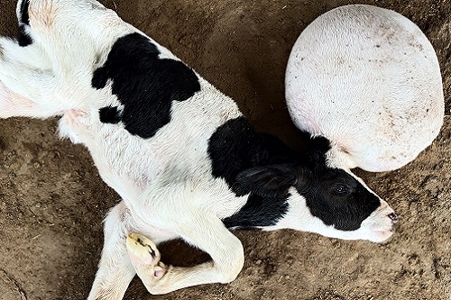
An animal owner from Village Bochasan, Taluka Borsad, Dist Anand (Gujarat) registered a visit in AMUL DAIRY that on his dairy farm a new borne male calf has an abnormal large round pot like growth on its head. On clinical examination, the live calf was in lateral recumbency, showing the symptoms of the abnormal dome-shaped growth on skull, weakness, poor suckling reflex, head tremors and convulsions (Fig. 1). It could not coordinate its movements to stand or walk at the time of examination. Physical examination revealed that frontal bones in the area of swelling were not formed and under the skin a fluid filled cavity was found. From the case history and clinical examination it was confirmed that the calf had congenital hydrocephalus. With the consent of owner, it was decided to operate and treat the patient.
Surgical Management and Discussion
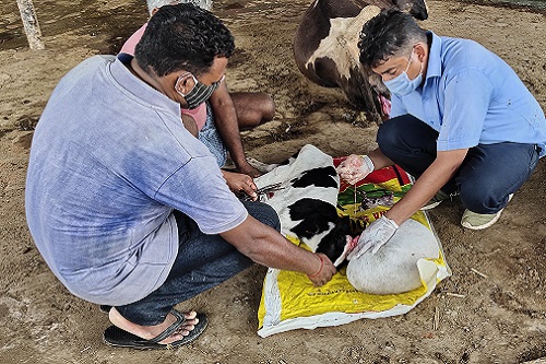
The site at the base of swelling was thoroughly cleaned & shaved. After scrubbing & preparation of site, 2% lignocaine hydrochloride was infiltrated around the swollen part of the head for achieving the local analgesia. Finally, the site was scrubbed and painted with povidone-iodine. A circular stab incision was given around the swelling. A small stab incision was made on the swelling to drain our the fluid slowly and to provide space at the base for further surgery. The whole mass was then removed by taking deep incision (Fig. 2). Then the skin edges were apposed in horizontal mattress pattern with non-absorbable cotton thread.
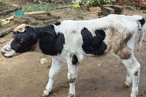
The calf was treated with Inj. DNS 450 ml slow i/v drip, and Inj. Oxy LA 3 ml, Inj. Meloxicam 1.5 ml and Inj. Dexamethasone 1 ml i/m. Antiseptic dressing with betadiane of suture line was done daily for 10 days post-operatively. The calf started suckling and moving normally within 6 hrs of surgery. After 12 days, the sutures were removed and the animal showed uneventful recovery with normal healing of suture line. The animal was normal healthy with usual behavior till 2 months post-operatively as per owners history & self observation (Fig 3).
The present findings of external hydrocephalus confirmed the observations of Sharma et al. (2015) and Saini et al. (2019), who also recorded very similar type of congenital hydrocephalus in buffalo-calf and cow-calf, respectively, with successful surgical management. According to Sastry (1971) the external hydrocephalus results from either too much fluid formed and not drained rapidly by the arachnoid villi or due to a hindrance to the drainage of a normally produced fluid during intrauterine life. The condition appears as a flaccid liquid-filled sac covered with skin and contains clear serous fluid. Congenital hydrocephalus is seen sporadically in all large animal species, although it is relatively common in calves, and has been described in cattle (Dhami et al., 2007; Saini et al., 2019, Borkhatariya et al., 2020), buffalo (Sharma et al., 2015) and camel (Abubakr et al., 1998). The calf in the present case improved gradually following surgical treatment, and showed ability to behave like a normal calf. Moreover, no history of convulsion was recorded thereafter. The findings regarding poorly developed thinned frontal bones of the skull allowing accumulation of large volume of fluid externally in skin in dome-shape with signs of convulsion in the present case concurred with the observations of Whitlock et al.(2008), Mausumi et al. (2014) and Saini et al. (2019). The literature available so far revealed that the life span of the affected newborn was very less, but in this case, the calf was leading a normal life after 2 months of surgical treatment.
In this communication, a rare case of external hydrocephalus of calf in a pleuriparous HF crossbred cow and its successful surgical management has been put on record.
Acknowledgment
We acknowledge CEO, ARDA, and MD, Amul for providing necessary facilities and support for mobile clinical services.
Conflict of Interest: None.
References
- Abubakr, M.I., Ahmed, A.A., Nayel, N.M., & Fadlalla, M.E. (1998). Hydrocephalus in a newborn male camel calf. Journal of Camel Practice and Research, 5, 309.
- Borakhatariya, D.N., Raval, R.J., Vala, K.B., Chavda, B.P., & Prajapati, S.G. (2020). Congenital hydrocephalic monstrosity in an indigenous Gir calf: A case report. The Indian Journal of Veterinary Science & Biotechnology, 16(1), 69-70.
- Dhami, A.J, Rabari, B.S., & Parmar, B.C. (2007). Dystocia due to hydrocephalic fetus in a crossbred cow. Indian Journal of Field Veterinarians, 2(3), 47-49.
- Long (2001). Abnormal development of the conceptus and its consequences. In: Arthur’s. Veterinary Reproduction and Obstetrics. 8th W.B. Sounders Co., Philadelphia, p.141.
- Mausumi, N., Subhasis, N., Upadhyay, S.N., & Hazra, J. (2014). A case of congenital hydrocephalus in a calf and its successful recovery. Exploratory Animal Medicine Research, 4(2), 253-256.
- Saini, R., Sharma, S.K., Aziz, P.R., & Pooniya, R. (2019). Congenital hydrocephalus in a calf and its surgical management: A case report. The Indian Journal of Veterinary Science & Biotechnology, 15(1), 81-82.
- Sastry, G.A. (1971). Veterinary Pathology. 3rd edn, CBS Publishers and Distributors, New Delhi, India, pp. 345.
- Sharda, R., & Ingole, S.P. (2002). Congenital bilateral hydrocephalus in Jersey cow calf. A case report. Indian Veterinary Journal, 79, 965-966.
- Sharma, S.K., Monika Joshi, Khosa, J.S., & Diler Singh (2015). An unusual case of dystocia due to hydrocephalic monster in a buffalo. International Journal of Science Environment and Technology, 4(2), 300-304.
- Whitlock, B.K., Kaiser, L., & Maxwell, H.S. (2008). Heritable bovine fetal abnormalities. Theriogenology, 70, 535-549.
| The content of the articles are accurate and true to the best of the author’s knowledge. It is not meant to substitute for diagnosis, prognosis, treatment, prescription, or formal and individualized advice from a veterinary medical professional. Animals exhibiting signs and symptoms of distress should be seen by a veterinarian immediately. |


this is not hydrocephalus, this is meningocele/meningo-encephalocele.
the surgery of the really hydrocephalus is by passing a tube from the cranium to the peritoneal cavity for absorption, what you have done is the surgery of meningocele