Lumpy skin disease, skin of cow showing ballooning and degeneration of prickle cell layer_edited
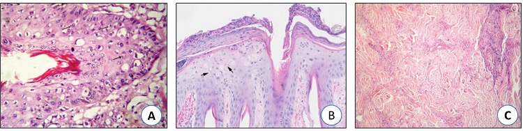
Fig. 4. A) Lumpy skin disease, skin of cow showing ballooning and degeneration of prickle cell layer with scattered intracytoplasmic inclusion bodies (arrows), H and E,×1200. B) The arrows point to eosinophilic intracytoplasmic inclusion bodies present in the keratinocytes. C) Lumpy skin disease, subcutaneous muscle of cow showing sever coagulative necrosis and calcification, H and E,×1200. (Neamat-Allah et al., 2015).
Be the first to comment