Abstract
This study discuss about the clinical management of lumpy skin disease (LSD) in a calf. A four months old calf having body weight of approximately 60kg was presented to Mobile Veterinary Clinics, Santipur Block (W.B.) with a history of nodular growth on different part of the body, reduced appetite, emaciation, nasal discharge and lameness. Clinical examination revealed that there were high body temperature (105°F), presence of various size of nodules on all over the body, particularly at neck region. Depending upon the history and clinical examination the case was diagnosed as lumpy skin disease (LSD) and accordingly therapy was given. For medical intervention Gentamicin (@5mg/kg B.Wt.) intramuscularly, Meloxicum (@0.2mg/kg B.Wt.) intramuscularly, antihistamine, multivitamins and antiseptic dressing of the lesions were given. Animal respond to the treatment and complete recovery was reported within 21 days.
Keywords: LSD, calf, clinical management
Introduction
Lumpy skin disease (LSD) is a transboundary, infectious. Eruptive, occasionally fatal disease of cattle and buffalo (Abdulqa et al., 2016) caused by Capripox virus from the poxviridae family (Ochwa et al., 2019). Sometimes the prototype strain is also termed as Neething virus (Gibbs, 2013; Abdulqa et al., 2016). Lumpy skin disease (LSD) is characterized by development of different sized nodular skin lesion including fever (Ochwa et al., 2019). The disease is transmitted mechanically by arthropod vectors (Mulatu and Feyisa. 2018). The virus is highly host specific and does not have zoonotic aspect. There is no sex and age predilection (Singh, 2019). Lumpy skin disease (LSD) first reported in 1929 at Zambia (Ochwa et al., 2019). But during the past few years this disease has spread through the Middle East into Southest Europe, Southest Russia and Western Asia (Singh, 2019). After the monsoon in India, humidity become very high during moist weather which is directly related with vector abundance (Gari et al., 2011; Mulatu and Feyisa. 2018). Morbidity of this disease are greatly varies ranges from 3 to 85% in different epizootic areas. In endemic areas morbidity is around 10% and mortality varies between 1-3% but in the condition of outbreak it may reaches up to 40% (Ochwa et al., 2019). This is an economically important disease of cattle and buffalo causes chronic debility in the infected animal, severe or permanent damage of hides due to skin nodules, severe emaciation, marked decreased of milk production and in some cases death of the infected animal (Zeynalova et al., 2016). The treatment of LSD is supportive to prevent the secondary bacterial infection with antibiotics (Feyisa, 2018) and in dressing of the lesions to prevent fly strike (Singh, 2019).
History & Observation
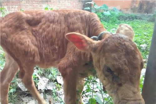
A four months old local breed calf was presented to Mobile Veterinary Clinics, Santipur Block (West Bengal) with the history of nodular growth on different body parts along with reduced feed intake. On physical examination animal was revealed emaciated, depressed and lethargic. And on clinical examination there was high fever (105°C), increased heart rate and respiratory rate along with swollen lymph nodes. Closed examination revealed there was different sized nodules on all over the body particularly at the neck region (Fig 1). The nodules are randomly distributed and some nodules were fused to performed larger nodules. The animal show lameness due to presence of nodules on the hind limbs.
Diagnosis & Treatment
In the countries where the disease is endemic, diagnosis is done only on the basis of history, observation, clinical findings and nodular lesions detected by the experienced veterinarians. Here on the basis of history, observation and clinical examination the presented case was diagnosed as lumpy skin disease (LSD).
The calf was treated with supportive therapy to prevent the secondary bacterial infections with antibiotics and dressing of the lesions. For medical intervention Gentamycin (JANTA®, @5 mg/kg B. Wt.) intramuscularly for five successive days, chlopheniramine maleate (CPM®) intramuscularly for three consecutive days, meloxicum (Melonex® @0.2 mg/kg B. Wt.) intramuscularly for two days and multivitamin (TRIBIVET®) 3ml intramuscularly for three days were given. The body temperature was dropped after 48 hours and sustained feed intake. Antibiotic lotion (Liq. Betadine) to clean the skin lesions, a fly repellent and healing cream containing gamma benzene hexachloride were given to apply topically twice daily until the nodules were disappeared. Within three weeks of the treatment the nodules were almost disappeared with the scars on skin and reported that calf was fully recovered.
Discussion
The principle manifestation of LSD are small to large sized nodular growth on the different body pats involving head, neck, limbs, genitalia etc. The regional lymph nodes are enlarged which are easily palpable. The affected animals may show high rise of body temperature, anorexia, depression and lameness.
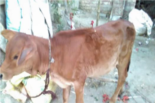
LSD is not associated with high mortalities (1-3%), but the economic losses is higher due to decrease feed intake, which causes chronic debility, reduced milk production, infertility, damage the quality of hide caused by nodular eruption on all over the body. Therefore antimicrobial and antihistaminic drugs are given for combat the skin infection and respiratory tract infection. In present case, antibiotic Gentamycin and antihistaminic Chlorpheniramine maleate were given to prevent the secondary bacterial infection and respiratory infection respectively. NSAID (nonsteroidal anti-inflammatory drug) meloxicum was given to reduce the body temperature. As animal was emaciated thus multivitamin also given to stabilized the health of the animal. Antiseptic lotion Betadine and fly repellent cream were given to improve the skin lesion. Within 21 days of above treatments the calf was completely recovered from the presented condition (Fig 2).
Conclusion
Lumpy Skin Disease (LSD) is an economically important viral disease of cattle and buffalo characterized by nodular skin lesions on different body parts. Consequence of this disease is very important on the aspect of hide industry as it reduces the hide quality. Dairy industry also affected badly as it causes marked decreased in milk production. As LSD is a viral disease, no particular curative drug is there, but supportive therapy with antibiotics, antihistaminic, anti-inflammatory and multivitamins can give to avoid the further complication and saving the life.
References
- Abdulqa, H.Y., Rahman, H.S. Dyary, H.O. and Othman, H.H. 2016. Lumpy Skin Disease. Reproductive Immunol Open Acc. 1(4): 25.
- Ganguly, S. and Yaday, S. 2016. Lumpy Skin Disease: A Brief Overview on the Trans boundary Animal Disease. Int. J. Phar. & Biochem. Res. 3(6): 1-2.
- Mulatu, E. and Feyisa, A. 2018. Review: Lumpy Skin Disease. J. Vet. Sci. & Tech. 9(3): 535.
- Ochwa, S., Vanderwaal, K., Munsey, A., Nkamwesiga, J., Nsekezi, C., Auma, E. and Mwiine, F.N. 2019. Seroprevalence and risk factors for lumpy skin disease virus seropositivity in cattle in Uganda. BMC Vet. Res. 15(1): 236.
- Gibbs, P. 2013. Merck Veterinary Website http//www.merckvetmanual.com
- Feyisa, A.F. 2018. A Case Report on Clinical Management of Lumpy Skin Disease in Bull. J. Vet. Sci. Technol. 9(3): 538.
- Gari, G., Bonnet, P., Roger, F. and Waret-Szkuta, A. 2011. Epidemiological aspects and financial impact of lumpy skin disease in Ethiopia. Prev. Vet. Med. 102: 274-283.
- Zeyanalova, S., Asadov, K., Guliyev, F., Vatani, M. and Aliyev, V. 2016. Epizootology and molecular diagnosis of lumpy skin disease among livestock in Azerbaijan. Front. Microbiol. 7: 1022.
- Singh, R. 2019. Outbreak of Lumpy Skin Disease (LSD) in Cattle in Chhotanagpur Platue Region (India). www.pashudhanpraharee.com
| The content of the articles are accurate and true to the best of the author’s knowledge. It is not meant to substitute for diagnosis, prognosis, treatment, prescription, or formal and individualized advice from a veterinary medical professional. Animals exhibiting signs and symptoms of distress should be seen by a veterinarian immediately. |



Be the first to comment