This article is beneficial for both veterinarian and for livestock owners, because if farmer can judge the abnormalities in behaviour of animal, only then he/she will be able to recognise the sick animal and be able to call the doctor for treatment. And I will tell very practical clinical signs in different species of dairy animals for veterinarian by seeing those clinical signs he/she can collect the blood sample for laboratory diagnosis, and be able to give some first aid until the lab report is not received. Article is on haemoprotozoan (blood parasites) of dairy animals (cow, & buffalo) These are of two types:
- Intra-erythrocytic ,or live in RBC’S eg: Anaplasma, Theileria or Babesia.
- Extra-erythrocytic or live outside the RBC’S eg: Trypanosomes
Transmission
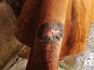
Also known as tick born diseases, haemoparasites are transmitted from one animal to other animals through ticks, flies. Also transmitted mechanically by blood transfusion, infected surgical instruments or when we do not change the needle during vaccination.
Clinical signs
These haemoprotozoan diseases are seasonal; commonly affect the animals in warm and humid weather when there is lot of tick population.
Common clinical sign which are present in all hemoprotozoan
- High fever
- Anaemia
- History of ticks on animal body.
Apart from common clinical signs there are certain specific signs of each haemoprotozoan disease
Intra- erythrocytic haemoprotozoan
Anaplasmosis
Also know as gall sickness caused by Anaplasma marginale (more pathogenic) and Anaplasma centrale (less pathogenic). The clinical signs are usually very mild in calves below 6 month of age,cattle upto 3 years develop typical and fatal disease, above 3 years disease is per acute and more fatal , infected cattle became carrier of anaplasmosis but are generally immune for further infection. (Handbook on Infectious Animal Diseases Shukriti Sharma, Vishal Mahajan & Kulbir Singh Sandhu 2008 Edition page no 103 )
Clinical signs: Very typical clinical signs of Anaplasmosis are:
- Difficulty in breathing due to exertion, mild coughing and there is open mouth breathing, this is due to anaemia.
- All mucous membranes become pale or yellow rather than healthy pink, we can check from eyes and vulva mucous membrane.
- Constipated faeces due to intestinal stasis and the faeces are coated with mucous membrane
- Staggering gait of animal
- Fever is recurrent (it is not throughout the day), either in the morning or in the evening with rapid bounding pulse.
Post mortem lesion or findings: when we open the body of dead animal there is jaundiced appearance of all visceral organs, enlarged gall bladder, and fatty liver.
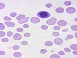
Babesiosis or Red water disease
It is vector born disease. Major vector for Babesia bigemina is Rihipicephalus microplus. Haemoprotozoan are pear shaped piroplasm joined at acute angle and present in the RBC’S.
Clinical signs: Apart from high fever and anaemia there is destruction of large number erythrocytes by piroplasm of Babesia resulting in haemoglobinemia and consequently haemoglobinuria,increased respiratory rate, may be diarrhoea or constipation pregnant animal my abort and death due the heavy loss of blood .
Post mortem lesions or findings: Most diagnostic lesion is urinary bladder filled with coffee coloured urine, gall bladder filled with thick granular bile.
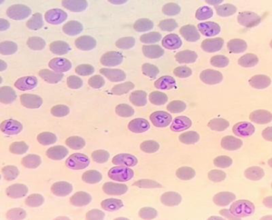
Theileriasis in dairy animals
It is also a tick born disease of cattle characterised by high fever and anaemia. Theileria anulata and Theileria parva are two organism causing disease. Theileria ovis is causing Theileriasis in goats. Theileriasis is mainly disease of exotic cattle.
Transmission: Rhipicephalus spp. of tick is responsible for transmission of disease from one animal to other animal. The mechanical transmission is not of much importance. After the bite the schizoints move into the lymphocytes and cause lymphadenopathy and then piroplasm of Theileria in erythrocytes.
Clinical signs: Lymphadenopathy, (inflammation of parotid lymph node in calves along with lacrimation, if not taken care leads to corneal opacity, and there is diarrhoea having blood . Inflammation of pre scapular and pre femoral lymph node along with diarrhoea having blood in adult animals is very typical clinical sign of subclinical Theileriasis. There is difficult respiration in animals (this is due to cystic mediastinal lymph node located in between the lung lobes). In clinical Theileriasis there is fever and anaemia, recumbency followed by death .There is cerebral form of Theileriasis in which animals exhibit circular movements, head pressing, salivation.
Post-mortem lesions and findings: very clear sign is cystic mediastinal lymph node and punched necrotic ulcers in the abomasum.
Extra erythrocytic protozoan
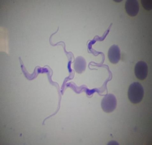
Trypanosomasis
It is also a vector born disease of dairy animals .Caused by Trypanosome evansi. Protozoan is present outside the RBC’S. Incidence of trypanosomiasis is high in rainy season due to large number of biting flies.
Transmission: Trypanosoma evansi is transmitted by biting flies/Tabanus/Glossina flies.
Clinical signs: In per acute form of disease animal dies after showing convulsions. (this is because of hypoglycaemia) high fever ,normocytic normochromic anaemia due to destruction of erythrocytes by flagellar action of parasites (Handbook of infectious animal diseases by Shukriti Sharma, Vishal Mahajan and K.S Sandhu 2008 edition page no 273), head pressing , intermittent fever, gazing of eyes, frequent micturition, corneal opacity unilateral or bilateral ,staggering gate and circling movement, wasting of muscles .
Post-mortem lesions and findings: all organs are anaemic, serous fluid in pericardial sac and abdominal cavity.
Diagnosis of haemoprotozoan diseases
Diagnosis of haemoprotozan diseases is based on clinical signs and laboratory diagnosis
|
Clinical signs |
Disease |
Laboratory diagnosis |
|
Anaemia, recurrent fever, jaundice like condition, difficult respiration on exertion, intestinal stasis, staggering gait etc. |
Anaplasmosis |
Under microscope solid dots of anaplasma marginale on the margin of the RBC ‘S, there is lymphocytosis with Eiosinophelia. CBC shows decrease in haemoglobin, low RBC’S count. |
|
Coffee coloured urine, high fever, high degree of anaemia |
Babesiosis |
Pear shaped organisms Babesia bigemina in RBC’S which are joined at acute angle. CBC shows, low haemoglobin and lymphocytosis withEiosiniphelia. |
|
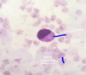
Initially only inflamed lymph nodes, lacrimation and diarrhoea having blood. Later on there is fever, difficult respiration, nervous signs like head pressing and circling movement. |
Sub clinical Theileriasis and clinical Theileriasis |
In sub clinical form there is koch’s blue bodies in lymphocytes (lymph node aspirate). In clinical Theileriasis there are piroplasms Thrileria anulata in RBC’S (signet ring like) CBC shows low haemoglobin in clinical Theileriasis, lymphocytosis and Eiosinophelia. |
|
High fever, head pressing, wide open eyes, paralysis of upper eye lid, hides bound condition. |
Trypanosomiasis |
Trypanosoma are present extracellularly. There is normochromic noromocytic anaemia, there is hypoglycaemia in blood. |
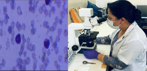
Precautions while collecting samples for laboratory diagnosis
- Always collect blood sample when there is fever
- In case of Trypanosomias is always give dextrose saline to animal solution before collecting the sample, and collect blood from ear vein.
- For the diagnosis of Theileriasis collect lymph node aspirate ( hold the lymph node ,inject 4-5 ml of normal saline solution ,do not withdraw the needle ,just massage the lymph node and try to retract back the injected saline solution. Make on the spot smear of this lymph node aspirate, dry it and fix it with methanol. Or you can send aspirate in syringe as early as possible to laboratory.
- Send blood in EDTA vacutanior. Try to send as early as possible.
Few tips for laboratory person
- Mix the blood before making smear.
- Try to make very thin smear.
- Always airs dry the blood smear,
- Fix the slide properly.
- Use distilled water for diluting Giemsa stain.
- Field A & Field B stain are very good for staining slides for haemoprotozoan.
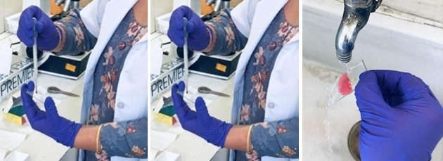
Take a small drop of blood ,put on grease free slide ,make thin smear ,dry in air ,put leishman’s stain for 1 min,wash the slide,put feild B stain for 30 sec, wash with water,put feild A stain for 1 min. Wash with water and dry in air . Examine the slide under 100x power of microscope .
Diffrential diagnosis
- From all diseases having high fever and anaemia.
- When there are nervous signs in case of Theileriasis, we should not confuse it with trypanosomiasis, confirmation by blood smear examination.
- Respiratory signs in Theileriasis should not be confuse with Heamorrhagic.
- After checking slide or after checking CBC it is very clear that there is anaemia with lymphocytocis in theileriasis but in case of H.S. there is neutrophelia.
- In Babesisosis haemoglobin urea, check the history of parturation (postparturient haemoglobinurea, respond to phosphorus therapy) and braken fern poisoning( history of ingestion of fern), leptospirosis confermation by PCR .
Treatment: It is strongly recommended to all farmers and veterinarians, never treat animals those are suspected for haemoprotozoan without laboratory diagnosis. Always try to give specific treatment to avoid drug resistance and burden on the pocket of the livestok owner. Sometime there is mix infection, like anaplasmosis and theileriasis most of the time ,but from clinical signs we are not able to judge clearly, which treatment is to be given. After taking blood sample we can give suportive treatment like, antipyretic ,dextrose saline etc. After looking the laboratory report specific treatment can be started
Anaplasmosis
Injection oxytetracycline 100ml in normal saline i/v for three days. If there is reoccurance or carrior state of disease then give oxytetracycline LA 20 mg /kg body wt i/m. repeat after 1 wk. Imidocarb is highly effective against anaplasma marginale a single injection @1.5 mg/kg s/c (The Merck Veterinary Manual 8th Edition Page no 23) suportive treatment with antipyretic, haemetinic syrup .
Theileriasis
Subclinical theileriasis: When blood picture shows only koch’s blue bodies in lymphoblasts and cbc shows lymphocytosis and there is no fever and anaemia we should treat the animal with schizonticidal drug lactate salt of halofuginone @1.2-2.0 mg/kg body weight orally for two days , or we can give injection of oxytetracycline –LA @20 mg /kg body weight i/m. (Handbook of infectious animal diseases by Shukriti Sharma ,Vishal Mahajan and K.S Sandhu 2008 edition page no260-261)
Clinical Theileriasis: when there is high fever with anaemia and blood pictuer shows piroplasms of Theileria anulata we should traeat the animal with buparvaquone(butalex) @2.5 mg/kg body weight i/m repeat after 48 hours. Suportive therapy is must like antipyretic and haematinics.
Trypanosomiasis
Many drugs are used to treat trypanosomiasis in dairy animals. Quinapyramine sulfate @ 5 mg/kg body weight s/c is used for treatment.and combination of quinapyramine sulfate and chloride are used for prophylactic and treatment @7.4 mg/kg body wt. (Handbook of infectious animal diseases by Shukriti Sharma ,Vishal Mahajan and K.S Sandhu 2008 edition page no 275). There is hypoglycaemia in animals so give dextrose saline i/v injections and other suportive treatment
Babesiosis
Injection diminazene aceturate is given i/m @ 3-5 mg/kg body weight. (Injection imidocarb dipropionate s/c injection @1.2 mg/kg body weight (The Merck Veterinary Manual 8th Edition Page no.25) suportive treatment with antipyretic and haemup syrups .
Controle of haemoprotozoan
Effective control of haemoprotozoan diseases is with control of ticks both on animals and in sheds , 80% of ticks live in sheds in groves of walls and floor of sheds and 20% of ticks on animal body . to kill ticks on body of animals apply drugs like amitraj pour on or butox. we can dipp the animal also when more number of ticks on the body of animals . we can also use injection ivermectin @ dose of 1 ml /33 kg body weigt s/c.
For control of ticks in sheds
1. Burning of sheds with hot air. 2. spray with malathion 0.5%.
For control of flies like tabnid flies
These flies develop in warm and damp places, therefore spray with kerosene oil or turpentine oil to kill the aquatic larvae. The manure should be frequently removed from sheds.
All slides are prepaired, examined and diagnosed by Dr Parvinder kaur, Veterinary Officer NRDDL, Jalandhar Punjab
Refrence:
- The Merck Veterinary Manual 8th Edition .
- Handbook of Infectious Animal Diseases by Shukriti Sharma ,Vishal Mahajan and K.S Sandhu 2008
|
The content of the articles are accurate and true to the best of the author’s knowledge. It is not meant to substitute for diagnosis, prognosis, treatment, prescription, or formal and individualized advice from a veterinary medical professional. Animals exhibiting signs and symptoms of distress should be seen by a veterinarian immediately. |

Excellent contribution by field vet for field 3
Very impressive and informative !!
Well done Dr sahiba !!
very informative article written in simple language and nicely explained by clear microscopic slides…
Very clinically relevant artilce …can I get pdf version…thanks for sharing knowledge
It’s very Informative clinical article for teaching Institute, field veterinarian…
Thanks
very good work dr sb
Very informative
Very knowledgable article and very nicely written.
As well knowledge for us and thanks for explain easly way
Very good Dr sab
Hi dr
Well said and it’s great you have written article.
Your knowledge and YouTube debate we learn and experience in our pig farm.
I’m harvir from Noida
Regards
Very good
Nice initiative, thanks.
Very informative
An informative article from field point of view
Very simple and informative
Excellent work by Dr Parvinder Kaur Lubana
Very informative n well explained Dr saab
Impressive⭐⭐⭐⭐⭐
Very informative
Very informative dr saab.
The article could be a ready reckoner for field vets. After reading this article one is encouraged to draw blood samples.
Very well written sir..also language is very easy to understand
Informative and clinically importance
Very quick guide, refreshing. Practical, after reading I come to know about importance of fomite spread. As HS vaccination is about to arrive, we should use separate syringes n needles to avoid fomite spread. Very clear slides are shown. This also shows importance of tetracycline group even today. A very good initiative. Also I would suggest regular training of field vets..
Beautifully Written in Simple n understandiable language which can be easily understand by field.
Please continue writing such articles
Beutifully Explained in Simple and easily understandable languange …could be helpful for field people..
Please continue writing Such articles.
Thanks
Best Wishes
Superb Write up ..7 Star.Very useful information for field vets.Takes you back to veterinary schools.
That’s great,… write up, pictures, all superb.Very informative.
Rating ⭐⭐⭐⭐⭐
A well explained informative article.
👍👍
Highly informative and compiled knowledge .
Best In simple to understand
Elaborated with best slides
Very nice article. Informative and educative.
Excellent very educative and informative combined nicely with theoretical knowledge and pictorial representations 👍🏻
Excellent work Dr Parvinder! Keep it up.
Excellent information given by author .very useful for field activities
Very useful guideline for Vets on field
Really imperative & beneficial too
Good Job done, nicely written.
Very simple to understand the slides and article . Great efforts put by Dr Parminder to make it so interesting and informative to read . Really blessed to have such gems in our department.
Excellent! Never seen such a beautiful, educative and beneficial to all , article. Great 👍 . 🌟 ⭐️ ⭐️ ⭐️ ⭐️ Rating
Excellent! Never seen such a beautiful, educative and beneficial to all , article. dr Parvinder ji has won Best Writer’s Award too ( PASHUDHAN PUNJAB – official magazine of Department Punjab ) . She has God Gifted quailities ont only in writing scientific articles but as a person too . Great 👍 . 🌟 ⭐️ ⭐️ ⭐️ ⭐️ Rating