Introduction
Urolithiasis describes the concretion of urinary calculi or organic compound, which may lodge any where in urinary system but most frequently at the distal end of the sigmoid flexure in ruminants and causes subsequent urine flow obstruction . Occurrence of urolithiasis is significantly more common in male ruminants compared to females due to their anatomical conformation of the urethral tract. The female has short ,wide and straight urethra while the male has long ,narrow and tortuous which makes them more prone to urethral obstruction, particularly distal aspect of the sigmoid flexure in bovines and urethral process in sheep and goats. Early castration of male might be the reason because it causes hypoplasia of urethra and leading to reduction in bore size of urethra. The decreased urethral orifice is a major predisposing factor for obstructive urolithiasis. In addition ,calculi formation usually results from a combination of nutritional, physiological, geographical, seasonal, age, sex and management factors. Treatment of obstructive urolithiasis is definitely surgical ,once the obstruction is complete. Removal of calculi may be by direct or indirectly bypassing the obstruction . Surgical tube cystostomy is the most commonly used treatment for long term management of urolithiasis in animals. It redirects the urine through a catheter placed from urinary bladder and existing through the abdominal wall. The present case study described the clinical signs and surgical management of obstructive urolithiasis.
History
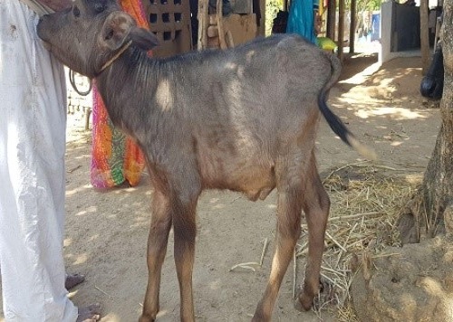
A case of 3 months old buffalo male calf referred to me having complete retention of urine since 2 days at village Borsad .
Symptoms
Clinical signs of the animals with an intact bladder were complete anorexia or inappetence, stranguria or anuria, reluctant to walk and frequent attempt to urination due to partial or complete obstruction. But in this case on physical examination of the calf , found bilateral distension of lower abdomen and its a sign of ruptured bladder.
Surgical procedure
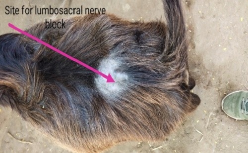
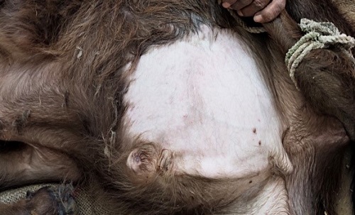
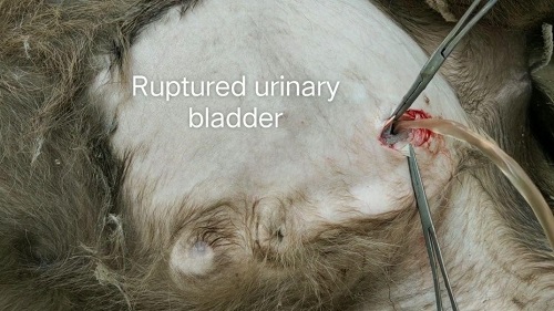
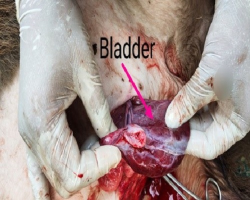
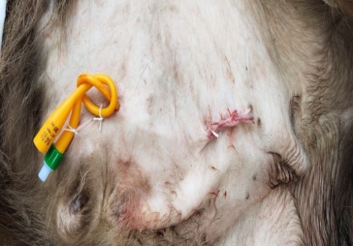
The calf was anaesthetised with local anaesthetic 2% lignocaine at lumbosacral junction by lumbosacral nerve block along with that line infiltration at surgical site. Animal was placed right lateral recumbency. Left side of the abdomen near the rudimentary teat the area was cleanly shaved and scrubbed with antiseptic solution. After scrubbing ,an incision was made nearly anterior to the rudimentary teat. Bladder was located after separating subcutaneous tissue and muscles by blunt incision ( Figure-2 ). The status of bladder was checked whether intact or ruptured . In this case bladder was found already ruptured as shown in Figure – 3. Here cystography done before using foleys catheter. After cystography subcutaneous tunnel was made parallel to the prepuce by passing straight forceps through the subcutaneous tissue at the end so that where skin was incised intended for the catheter outlet ( Figure-5 ). Foleys catheter was passed from outside to abdominal cavity where the catheter tip was held in stillette and directly stabbed the bladder & its bulb was inflated with the normal saline for fixation. Muscles and sub cutaneous tissue was sutured with catgut no.2.The foleys catheter was sutured at multiple sites on the ventral abdomen (Figure-5).
Postoperative treatment,3 to 4 times long acting oxytetracycline @ 20mg/kg b.wt & dexamithasone given on every 4th day along with ammonium chloride orally daily @ 200 mg/kg b.wt b.i.d.for minimum 15 to 20 days .Application of local antiseptic dressing with povidone iodine was advised for a week. The catheter was allowed to drain freely for minimum 10 to 15 days until normal urination resumed. In this case catheter was removed on 11th day of surgery after the confirmation of normal urination through the external urethral orifice .
Results
The calf is now recovered completely. Defecation, urination, feeding and all other activities are normal as per self observation & owners description.
|
The content of the articles are accurate and true to the best of the author’s knowledge. It is not meant to substitute for diagnosis, prognosis, treatment, prescription, or formal and individualized advice from a veterinary medical professional. Animals exhibiting signs and symptoms of distress should be seen by a veterinarian immediately. |
Be the first to comment