Abstract
This invention/methodology specifically describes a combination of medicines and time of administration, which is cheaper, economic, effective, quick recovery, less recurrence, viable technology, sensitive and specific as well. Use of three different medicines in combination has been described and all are cheaply available in the market. Use of this technology/methodology has opened a new vista to treat humpsore in endemic regions through this invention because this has improved health & wellbeing, milk yield, fertility rate, growth rate in heifers, and reduced stress and nuisance in the affected animals. Invented technique for treatment of humpsore by combined administration of different medicines, functionality and dosage of individual medicines have been standardised, does not require any specific dosages and equipments, reduces the time and cost of treatment. Therapeutic module of humpsore developed by this methodology could be used successfully for complete treatment with 100% effective, quick recovery and less recurrence as well.
Introduction
Livestock sector especially cattle enterprise constitutes an important part in wealth of a country as it provides milk, meat, manure, draft power and leather to the vast majority of the people throughout the world. Humpsore is caused by Stephanofilaria assamensis and spread or transmitted biologically by fly (Musca conducens). This disease widely affects the cattle, but also affects buffalo, goat, elephant, black rhinoceros and Nilgi in India. It is also considered as a zoonotic disease; however, its occurrence in humans is rare. It is a common disease in Eastern region of India mainly in Assam, Tripura, West Bengal, Odisha and Andaman and Nicobar Islands. It also occurs in Andhra Pradesh, Telangana, Bihar and Gujarat, besides Southeast Asian countries.
Symptoms
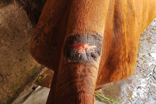
- Humpsore causes pruritis, damages in hair follicles and skin glands and It causes ulcerative nodular dermatitis, exudation, granulation, ulceration and incrustation depending on the stage of humpsore. Humpsore adversely affect the dairy cattle in endemic regions like Andaman and Nicobar Islands.
- Humpsore dermatitis is initially of serous exudate, crusts and papules; Chronicity leads to alopecia, skin thickening and hyperkeratosis.
- Humpsore dermatitis on the ventral midline consists initially of serous exudate, crusts and papules; Chronicity leads to alopecia, skin thickening and hyperkeratosis.
- Lesions are an extreme irritant and evidence of rubbing is present. Pruritus causes affected cows to attempt to scratch their bellies while they are partially recumbent. Cows may rise to their knees and rock the brisket and ventral abdomen fore and aft in an effort to relieve the itching sensation associated with dermatitis.
Predilection sites
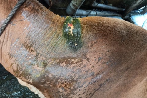
- Stephanofilariasis primarily affects the area in and around the hump (96.20%) but also affects eye inner canthus, horn, ear, legs, scrotum, udder, sternal area, back and ventral surface of the body, anterior and posterior to the navel and on the abdomen in cattle.
- In buffalo, humpsore is commonly affected in the horn (95.3%) followed by in ear (12.70%)
- Severity of the humpsore lesions varies with different predilection sites.
Factors influencing humpsore infection in ANI
- Humpsore size varies from a few cm to more than 30 cm in ANI because it has typical hot and humid climate throughout the year and rainfall extending over 8 months with higher relative humidity (average 80%) favours the un-interrupted growth of the parasites and flies.
- Thick vegetation with humid tropical climate of ANI favours the development of assamensis and fly.
- Prevalence of humpsore in ANI is ranged from 10.2 to 70.5% with highest incidence was in North and Middle (70.5%) Andamans and lowest incidence (10.2%) was reported in Little Andaman Island whereas moderate prevalence was recorded in Neil Island.
- Close contact of the animals and negligent management of cutaneous abrasions could be the cause of higher incidence of humpsore under farm conditions.
- Higher incidence of humpsore is commonly observed in exuberant high occupancy pastures with large quantities of wet faeces in hot and rainy seasons.
- Humpsore has showing higher prevalence rate in exotic and its crossbred cattle (20.17%) than in zebu (16.14%) cattle.
- Prevalence rate of humpsore was lower in light coloured cattle than in dark coloured cattle in zebu type.
- The age and sex of the host also influences this infection. Higher incidence was observed in female (85.70%) than in male (14.3%)
- Calves below one year of age has not been seen with humpsore lesions and the incidence is increased as age advances
- Higher incidence is in animals grazing on common pastures or near garbage areas (54.2%) than those reared in the backyards (24.4%)
- Further higher incidence is in street cattle than in organised farm cattle
- Humpsore is more common in the farm than villages as 31.63% of animals affected in farm than 26.00% in the villages.
- Humpsore is more common in dry season to reinfection and increased numbers of vector bites, thus apparently raising the proportion of infected animals than rainy season. Therefore, the treatment should be conducted once in six months or twice in a year in Andaman and Nicobar Islands
Adverse effect of humpsore
- Severe infections can cause severe stress to the affected animals which inturn affect production and reproduction performances.
- Humpsore predisposes severe economic losses to the livestock farmers.
- Humpsore affects the manual or mechanical milking and creates unclean milk.
- Humpsore affects the male and female animals. The affected male is unsuitable for draught and plough purposes and affected females lose their productivity, growth rate and fertility rate.
- Humpsore causes poor growth, delayed puberty in heifers and diminished milk yield, increased inter-calving interval period in milch cows.
- Humpsore is located mainly on or near the regions of the hump and neck, it causes extensive skin damage. The animal is having very ugly skin lesions with reduced value of the animals, causes huge economic loss to the farmers
- Humpsore results into reduced working capacity and increased hide damage. Damaged hides can be downgraded and even rejected at slaughter.
- Thus the farmers suffer heavy economic losses.
Pathology of humpsore
- A break in the skin epidermis is the pre-requisite condition for the subsequent development of humpsore
- Continuous ocular discharge from eye inner canthus and feeding of the discharge by flies
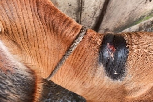
- Injury in the base of the horn and base of the ear by frequent rubbing of restraining rope
- Behind the poll with continuous rubbing of chain or raze mad to control the animal
- Middle and posterior neck occurred by frequent and continuous rubbing during ploughing
- Drawing cart and with oil or molasses machine
- Abdominal wall or other regions injured by rough floor of farm, barbed wire, during fighting with each other
- Injury on the hump/withers by manger under farm conditions
- By metallic rod or bamboo at the open place animals to prevent soiling of the food in the manger
- Most important clinical sign is intense pruritis with characteristics of rubbing of the sore with wall, pillar, trees or fencing leading to extensions of lesions which become central ulcerative or excoriation of the parts followed by formulation of a cauliflower like appearance due to enthusiastic granulation on the skin.
- At first, the small papules are formed and later on papules get ruptured leading to coalesce and form large lesion.
- The exudates get dried to form the crust followed by thickened skin.
- Ultimately there is loss of hairs, hyperkeratosis, ulceration and haemorrhage.
- The lesions attract large number of flies which lay eggs in them.
- Crows play an important role by causing a great deal damage through pecking at the wounds and keep them fresh, active and bleeding.
- Dyskeratosis is a disorganised arrangement of epidermal cells with mitotic figures leading to a certain degree of anaplasia in humpsore lesions.
- Filariids produce pathological effects in long-standing cases. The lesions become blackish during winter when it dried and again it turns out to be active in monsoon or rainy season.
- Secondary bacterial infection ((Staphylococcus aureous, Staphylococcus albus ) and/ or maggot infestation aggravate the situation.
- Musca flies become infects with microfilariae when they bite or feed on the wounds of cattle. Microfilariae develop to infective larvae (L3) inside the flies in about 3 weeks. The infected fly carries the microfilariae in proboscis and inoculates the microfilariae to healthy animals by biting or feeding in wound.
- In initial stages of wound, the skin is covered by grayish white crust which favour for stephanofilarial infection. The non-ulcerated lesions frequently become excoriated favouring further deposition of stephanofilarial larvae and gradually obtain the characters of ulcerated lesions resulting partial or completely devoid of hairs.
- The ulcerated lesions have central ulcers of various sizes and shapes surrounded by larger peripheral crusty areas in most of the lesions with irregular boundary.
- The discharge of the ulcers is pureblood, serum or blood mixed serum.
- The surface of the ulcers is frequently hemorrhagic and moist but dry surface covered by thin blackish or brownish crusts and a number of flies are seen feeding on the discharges.
- The colour of the crusty/scabby part is found to vary from grayish white to blackish and this part is raised from the ulcerated part or normal skin surface. The thickness of the crusty part of the lesions is up to 2.3 cm according to character and degree of crust formation, compared to the normal skin thickness of about 2.5 mm. The shape of the lesions is characteristically circular although roughly rectangular, triangular or irregular shaped lesions are common.
- Close contact of the animals and negligent management of cutaneous abrasions could be the cause of higher incidence of humpsore under farm conditions.
- Clinically two types of lesions are considered for descriptive and diagnostic purposes. Early lesions are smaller in size (3 to 4 cm in diameter) characterized by formation of thin, mildly granular crusts in most lesions and rarely laminated. The old lesions are larger in size and have coarsely granular crusts with many cracks and crevices with or without centrally located ulcer. Both types are partially or completely hairless.
- Humpsore lesions are again classified according to severity depending on amount of crusts, exudation, size of lesion and ulceration into three types such as mild, moderate and severe.
- Humpsore is more common in dry season (October to April) as at the beginning of rainy season (May to June) there is formation of small size crust form of filariasis and at the end of the rainy season (September), oozing is occurred.
- During the dry season, October onwards, the wound size becomes gradually increased and become active and increased oozing. Moreover, during the dry period, the fly population is also increased at highest level. Humpsore is very common and increased size during the dry season and consider the infections as seasonal.
Therapeutic approaches
- Completely approved treatments for stephanofilariasis are not available for cattle and buffaloes and different treatment protocols have varied success rate. The therapeutic approaches were assessed on the basis of skin healing and normal posture of the animal.
Table Existed therapeutic measures for Humpsore
| Sl. No. | Treatment Protocol | References |
| 1 | Surgical removal or cauterizing agents | Mishra (1969) |
| 2 | Ointment made from 64 preparations (conch shell ash, lead monoxide, sulphur, tobacco, etc.) | Hassan (1969) |
| 3 | Application of Petroleum jelly | Patnaik (1970) |
| 4 | Neguvon 6% liniment + Asuntol 6% dusting powder | Patnaik (1970) |
| 5 | Antimosan (s/c) + 1% gentian violet | Ahmed and Ali (1973) |
| 6 | Trichloropon applied with 6-10% petroleum jelly or castor oil | Rahman and Khaleque (1974) |
| 7 | 8% Trichlorophon + 4% sulphonamide ointment
(Healed within 20-26 days) |
Baki and Dewan (1975) |
| 8 | Parentral administration of 8% Trichlorphon (very effective) | Baki and Dewan (1975) |
| 9 | Antimony potassium tartrate + Phenothiazine (4-8% ointment) | Dutta and Hazarika (1976) |
| 10 | Tobacco ointment (80% of cases cured) | Dutta and Hazarika (1976) |
| 11 | External application of Antimony potassium tartrate | Dutta and Hazarika (1976) |
| 12 | Trichlorophon (6-10%) in petroleum jelly or castor oil (Cured within 7 days) | Rahman and Khaleque (1974) |
| 13 | Supona 20 and Sumithion @ 4% concentration (Toxic signs @ 6%) | Das et al. (1977) |
| 15 | Levamisole hydrochloride + blanket treatment | Rai et al. (1994) |
| 16 | Levamisole HCl + Zinc oxide ointment
(mild and moderate size healed within 5-13 days) |
Rai et al. (1994)
Rai and Ahlawat (1995) |
| Less than 2 cm diameter healed within 5-6 days | ||
| 2-5 cm diameter healed with 7-8 days | ||
| 5-8 cm diameter healed within 10-14 days | ||
| 8-12 cm diameter healed within 15-18 days | ||
| more than 12 cm diameter healed within 17-25 days | ||
| 17 | Ivermectin + Levamisole + Mastilep ointment | Choudhury and Das (2012) |
| 18 | Levamisole (3ml s/c) at lesion+ Dermocept ointment (healed in 11 days) | Phukan et al. (2005) |
| 19 | Ivermectin + Topicure spray
(healed within 21 days) |
Puttalakshmamma et al. (2012) |
| 20 | 15% Tobacco ointment | Al Masud et al. (2017) |
| 21 | Ivermectin + Zinc oxide ointment twice daily | Islam et al. (2018) |
Side effects or disadvantages of the existed therapeutic methods
Neguvon (6%), Nuvon (6%) and Dimecron (6%) were cured the humpsore disease within 20-25 days. However they act as the neurotoxin causes neuro-generative disorder and as a potent carcinogen causes malignancy. Not only that it also induces respiratory tract irritation, gastrointestinal disorders and CNS related problems such as tremors, disorientation, memory loss and cognitive dysfunction in short and long term condition. It also induces liver damage and diabetic disorders. On the other hand, Diethylcarbamazine citrate ointment showed poor results than in injectable form. DEC @ 20% solution and injection on day 0 and 10 (S/C) around the wound with daily application of zinc oxide ointment cured within 20-30 days depending upon the size with the limitation of only effective against adult stage of the organism. Similarly Levamisole HCl (1 ml/15-20 kg BW) as single deep I/M or S/C injection followed by daily zinc oxide ointment application cured in 8-20 days depending upon the size of the wounds. Small wound takes 8-10 days and larger wound takes 10-20 days. However it has contradiction like it is not approved in lactating animals. It should be used cautiously, if at all, in animals that are severely debilitated, or have significant renal or hepatic impairment. Use cautiously or, preferably, delay use in cattle that are stressed due to vaccination, dehorning or castration.
Innovative therapeutic methodology: Trimodel Therapy Module to Treat Humpsore
In human filarial diseases (Onchocerciasis and lymphatic filariasis), albendazole in combination with ivermectin, diethylcarbamazine (DEC) or levamisole is used to treat these filarial diseases. This treatment protocol includes use of microfilaricide (kill immature worms; such as DEC) and microfilaricide (kill adult worms; such as Ivermectin). This therapeutic protocol is modified and used in dairy animal to treat humpsore.
The treatment procedure is as follows:
- Humpsore is cleaned with a mild liquid soap to remove the dirt, antiseptic solution and crusts are removed.
- Un-ripened papaya (Carica papaya) juice was applied on the wound for about 30 min for first three days to digest the dead tissues.
- Application of antiseptic and fly repellent ointment daily to check bacterial infections and as a fly repellent for 30 days
- Injection of Ivermectin/Doramectin @ 200µg/kg body weight is done by subcutaneous/deep intramuscular route at fortnight interval for a month.
- Diethylcarbamazine 6 mg/ kg body weight was administered orally in morning daily for 30 days.
Papaya latex contains enzymes, papain, chymopapain, caricain, glycyl endopeptidase and papaya lipase, which have antiviral, antibacterial, antiprotozoal, antifungal, and anti-inflammatory and wound healing properties. It desloughs the necrotic tissue and prevents wound infection. Papaya is also used as an ulcer protector. Papain is a cysteine proteinase that digests necrotic tissue by liquefying eschar, thus facilitating the migration of viable cells from the wound edge into the wound cavity. Papain is also reducing the bacterial burden, decreasing exudates and increasing granulation tissue formation. Papain isolated from the latex of unripe papaya pulp is used in wound care and chronic skin ulcer therapy as it has anti-bacterial and fibrinolytic properties.
DEC is drug of choice to treat the filariasis. It kills both microfilariae and adult worms. Diethylcarbamazine is well absorbed following oral administration. It reaches all parts of the body within 25 minutes after its intake with peak plasma concentrations reached within 1-2 hours. The plasma half-life varies from 6.1 to 8.1 hours. The standard DEC treatment regime is 6 mg/Kg per day over a 10 to 20 day period in filariasis treatment. The elimination half life ranges from 10-12 hours. If the urine is alkalinized, renal excretion of unchanged drug is prevented and the half life of the drug increases as cattle is herbivores, hence half-life of medicine may be probably higher. It has an antagonistic action of the metabolic enzymes that metabolize arachidonic acid indicates minimizes the inflammatory condition. On the other side, DEC involves two processes as in first process, the filarial muscular activity decreases, and secondary to hyperpolarization of membranes by the piperazine moiety of DEC and in second process, the microfilarial surface membranes are altered by making them more susceptible to host defenses. In general, the filarial microfilaria reach peak in morning hours. Therefore, DEC was administered at the morning time and at 24 h interval to the cattle to treat humpsore for 20-30 days depending upon the degree of the humpsore.
Ivermectin is widely used anti-filarial drug and it is a macrocyclic lactone with broad spectrum activity on filarial parasites. Ivermectin interacts with postsynaptic glutamate-gated chloride channels (GluCl), causes an influx of Cl- ions through the cell membrane by activation of specific ivermectin-sensitive ion channels, resultant muscle hyperpolarization leads paralysis of the microfilarias. Ivermectin acts quickly on the microfilariae and with long-lasting effect, inhibits adult female worms to release additional microfilariae. Dermal microfilarial loads are generally decreased by 78% within two days and by some 98% within two weeks of Ivermectin treatment. Elimination half-life of ivermectin in cattle is 2.0, 3.7, and 8.3 days with aqueous, aqueous: glycerol-formal (50:50 v/v), and propylene glycol: glycerol-formal (60:40 v/v) formulations, respectively whereas its metabolites may persist for more days. Lowest levels of dermal microfilariae observed after this timeframe, it indicates that ivermectin affected microfilariae all are not killed in the first few days. This is supported by reports that microfilariae migrate into deeper dermal layers, sub-cutaneous fat, connective tissue and lymph nodes following administration of the ivermectin. Ivermectin actually interferes with the ability of microfilariae to evade the immune system, resulting in the host’s own immune response being able to overcome the immature worms and so kill those microfilariae. It is well known that ivermectin administered SC has persistent for 10-21 days. Therefore, we administered ivermectin at the interval of 15 days (day 0, 15 and 30) in the study to treat humpsore.
Commercial ointment has miticidal, antifungal, antiseptic, anti-inflammatory and fly repellent properties. It is used in skin lesions which are produced by a variety of agents including external irritants, burns, allergents, trauma and viral, bacterial, parasitic or fungal infections. In case, this commercial ointment is not available, we recommend the paste which is made up of 100 ml neem oil or coconut oil + 5 camphor + 50 g turmeric powder + 1 lemon. This ointment or paste needs to be applied for 30 days of treatment.
- Fliesdon’t like the smell or fumes from camphor
- A 2018 animal study found that an ointment containing camphor and coconut oil decreased the healingtime for second-degree burn woundsand was found to be more beneficial than using Vaseline.
- The curcumin found in turmeric can help wounds heal by decreasing inflammation and oxidation. It also lowers the response of your body to cutaneous wounds. This results in your wounds healing more quickly. Studies have found that turmeric can positively affect tissue and collagen as well.
- The lemon is also used for healing up the wounds, thanks to the great quantity of citric acid that it contains, as well as to stop hemorrhages.
Advantages of the Technology/methodology
No such type of the methodology/ Trimodel Therapy Module available in India or abroad to treat humpsore. In Andaman and Nicobar Islands, 30-40% animals suffered humpsore, out which, 70.5% in North and Middle Andaman, 10.2% in Little Andaman and remaining in South Andaman. Out of dairy cattle population, good population of animals remained infected with humpsore, which requires correct treatment at the right time. Hence the technology/methodology developed has profound economic significance.
- The treatment methodology is economical and methodology is viable. Treatment to the humpsore is ongoing and regular activity which is essential to enhance the economic status of farmers and increase the production and reproduction performances of the dairy cattle.
- This treatment methodology is cheaper, economic, effective, quick recovery, less recurrence, viable technology, sensitive and specific as well for cattle and buffaloes except the pregnant animals.
- The treatment methodology has improved milk yield (25-30%) and fertility rate (30-40%).
- The treatment methodology has reduced the stress level and nuisance due to humpsore (decreased malondialdehyde: 15-20% and cortisol: 20-25% and increased total antioxidant capacity: 5-10%, superoxide dismutase: 20-30%, catalase: 12-30%).
- This methodology has reduced the inter-calving interval and improved estrus induction rate and growth rate in heifers.
- No recurrence since one year.
Method of administration and medicine availability
It is not a complicated method of administration. Ointment or paste is applied externally, DEC is given orally and Ivermectin in subcutaneous or Doramectin in subcutaneous/deep intramuscular route. These medicines and ointment are available in commercial veterinary pharmacy. The cost of the therapy per animal is Rs.431/-, which is very cheaper.
Impact of the methodology/technology
The field application of the ICAR-CIARI technology has improved the milk yield (25-30%) and fertility rate (30-40%) and reduced the stress level and nuisance due to humpsore (decreased malondialdehyde: 15-20% and cortisol: 20-25% and increased total antioxidant capacity: 5-10%, superoxide dismutase: 20-30%, catalase: 12-30%) indicating that a significant increase in health and wellbeing, production and reproduction performances in dairy cattle. Considerable interest has been generated in this work and industries/ farmers/ organisations of eastern parts of India have shown their interest in the technology. Therefore, this technology needs to be propagated among a large population of consumers including farmers/dairy owners/field veterinarians/extension workers throughout the country.
Prevention and control
- Control and eradication of humpsore in cattle can be successfully achieved by targeting the life cycle of the fly, Musca conducens.
- Simultaneously, blanket treatment of affected animals during active phase of the parasite after cordoning the area
- Improvement in general cleanliness and hygiene to reduce the fly population
- Promptly attending to fresh injuries/cuts and concerted efforts to make the intermediate host sterile for parasite may be undertaken.
Fund Source
National Bank for Agricultural and Rural Development (NABARD), Port Blair, Andaman and Nicobar Islands sponsored in the form of the project entitled “Humpsore treatment by Trimodel therapy to improve the Production and Reproduction Performances of milch cows in South Andaman” for validation, popularization and propagation of this technology.
| The content of the articles are accurate and true to the best of the author’s knowledge. It is not meant to substitute for diagnosis, prognosis, treatment, prescription, or formal and individualized advice from a veterinary medical professional. Animals exhibiting signs and symptoms of distress should be seen by a veterinarian immediately. |


Be the first to comment