Introduction
Poisonous and venomous animals are abundant and include more than 400 species of venomous snakes, approximately 1500 species of toxic marine animals, and countless numbers of poisonous or venomous arthropods distributed widely throughout the world. Virtually every phylum of the animal kingdom is represented by species that produce toxins, termed zootoxins, in the form of either poisons or venoms. Poisons are toxins that are produced as secondary metabolic products that accumulate in the host animal tissue or that accumulate in tissues of predators after ingestion of toxin-bearing prey (e.g., ciguatoxin accumulation in predatory fish such as barracuda or jack). Not all exposures to toxic animals result in toxicoses, as variations in toxicity of zootoxins can occur because of a host and environmental factors such as age, sex, nutritional status, season, geographic location, and toxin composition. Similarly, target animals may vary in their susceptibility to zootoxins because of factors such as age, size, species, location and severity of bite/sting. Following are some zootoxin producing animals, along with their treatment, to which humans and animals are mostly exposed
1. Marine zootoxins
Marine zootoxins include toxins from dinoflagellates or algae that accumulate in the tissues of marine animals, e.g., domoic acid, gonyautoxins, ciguatera toxin. The toxin may be produced by the fish itself or by the marine plankton or algae consumed by the fish. Fish is the main source of proteins for inhabitants of the coastal areas and also for the animals in the form of fish meal in their feed. Fish poisoning (Ichthyotoxicosis) is more common in human beings than in animals. Some common ichthyotoxins have been discussed below-
Domoic Acid– Domoic acid is a heterocyclic amino acid neurotoxin produced by a variety of marine algae and with a structural similarity to kainic acid. The neurologic and behavioural effects of domoic acid are thought to be due to its mimicry of the neurotransmitter glutamate within the nervous system. Domoic acid toxicosis has been associated with gastrointestinal, cardiovascular, and neurologic dysfunction in many species. In humans, toxicosis associated with domoic acid has been characterized as predominantly gastrointestinal upset (vomiting, nausea, diarrhoea, abdominal discomfort/cramping). Neuropsychiatric effects are less common and include severe headache, confusion, disorientation, loss of short-term memory (inability to recall recent past), myoclonus, agitation, seizures, and coma. Clinical effects may persist for days to months after exposure, with some neurologic effects such as short-term memory loss persisting for years. Deaths are not common but may occur within days to months of exposure.
Ciguatera– Ciguatera fish poisoning results from the ingestion of fish (Moray eel) that have accumulated ciguatoxins produced by the dinoflagellate Gambierdiscus toxicus. Ciguatoxin is a neurotoxin that acts on voltage-sensitive sodium channels on excitatory membranes of nerves and muscles resulting in excess intestinal fluid secretion (diarrhoea, cramping), vomiting, autonomic dysfunction (hypotension, bradycardia), paraesthesia, muscle weakness, myalgia, arthralgia, fatigue, pruritus, and numbness of lips and/or limbs. The neuromuscular signs may persist for weeks to months, and in severe cases, death can occur.
Tetrodotoxin– Tetrodotoxin (TTX) is produced by a marine bacterium and accumulates in the tissues of a poisonous Pacific fish, the pufferfish. It is a selective voltage-dependent sodium channel blocker nonprotein toxin. The consumption of an organism containing TTX can cause neurological and gastrointestinal symptoms. TTX, widely distributed among marine as well as terrestrial animals, induces dangerous intoxications. This toxin is mainly isolated from the pufferfish’s skin, viscera, ovaries, and liver. It is a very potent toxin i.e., the intraperitoneal LD 50 value in mice is 8 µg/kg.
Saxitoxin– It is produced by Alexandrium spp. and related dinoflagellates and mainly reaches into the body after ingestion of shellfish. Saxitoxins exert their effect by binding directly to the voltage-dependent sodium channels in nerve and muscle cell membranes, interrupting nerve signal transmission and leading to paralysis.
In contrast to ciguatera poisoning, gastrointestinal symptoms are less prominent than neurologic manifestations. Circumoral paraesthesia and paraesthesia of the extremities usually appear within 1 hour of ingesting toxic shellfish, and they may be accompanied by ataxia, dysphagia, and changes in mental status. Hypertension may occur and corresponds to the extent of the ingested dose. In the most severe cases, patients may proceed to respiratory paralysis, usually within the first 24 hours of illness.
The lethal dose of saxitoxin in human beings is approximately 4 mg.
Treatment
No treatment modalities exist to prevent the effects of ichthyotoxins other than to remove the patient from the source of the toxin and provide symptomatic and supportive care.
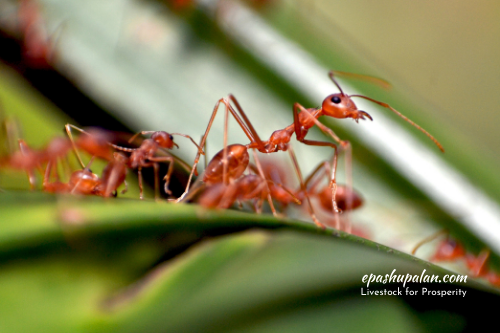
2. Zootoxins from Insects
Hymenoptera (bees, wasps and ants) are the most commonly reported insects involved in toxicosis. Hymenopteran venoms contain a mixture of proteins, peptides, and small organic molecules. Ant venom tends to have low protein content and is made up primarily of alkaloids, whereas bee venom has a much higher protein content. The toxicity of bee venom is heightened by the presence of hyaluronidases and phospholipases, which account for a majority of the allergic responses seen with bee envenomation. Melittin, a membrane-disruptive component of bee venom, can induce haemolysis, increase capillary blood flow, increase cell permeability, and enhance the spread of venom within the tissue. Apamin is a neurotoxic compound that blocks potassium channels and may be responsible for peripheral nerve dysfunction that occasionally occurs in humans after bee stings.
The severity of toxicity varies from individual to individual in different species of animals. Anaphylaxis and death from a single sting occur in hypersensitive animals. However, only mild to moderate inflammation and painful swelling are observed in others. Following a single insect bite, there is extreme serous exudation. However, in multiple bee stings, there is severe local inflammation and oedema at the site of sting, intense pain and pronounced excitement. In severe cases in horses, there may be diarrhoea, methemoglobinemia, bilirubinaemia, jaundice, haemoglobinuria, tachycardia, dyspnoea, and followed by death, though in rare cases.
Treatment
No specific antidote is available. Only symptomatic treatment needs to be given.
- Local application of a weak solution of ammonia and sodium bicarbonate.
- Nervine tonics and stimulants should be given, if there is prostration.
- Tracheotomy, if asphyxia is severe.
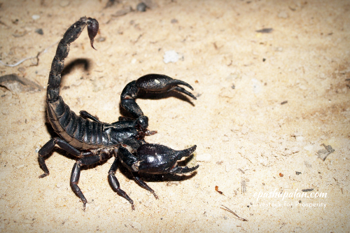
3. Zootoxins from Arachnida (Spiders and Scorpions)
Spiders: Numerous species of spiders have been implicated in bites of human beings and animals. These are- Black widow spider, brown widow spider, red-legged spider, brown recluse spider, etc. Among these, most of the reported cases are due to black widow spider (Latrodectus mactans) and brown recluse spider (Loxosceles reclusa).
Widow spiders have an oily venom composed of a mixture of neuroactive proteins, of which α-latrotoxin is the most potent and responsible for most of the signs and symptoms associated with envenomation. The binding of α-latrotoxin to neuronal receptors results in a massive release of acetylcholine (ACh) at the neuromuscular junction, resulting in profound muscle contraction. The symptoms of toxicosis relate to this neuromuscular activity and include muscle spasms, muscle pain, and abdominal rigidity. Hypertension, tachycardia, hyperesthesia, vomiting, and diarrhoea may also occur.
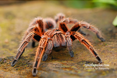
Recluse spider venom contains a variety of necrotizing enzymes, of which sphingomyelinase D is the most important. The effects of Loxosceles spp. toxin is local in nature, causing a spreading necrotic area that may ultimately encompass a large tissue area. Systemic reactions including haemolysis, dyspnoea, and coma, can occur but are quite rare.
Scorpions: In many parts of the world, scorpion envenomation is a significant public health concern. Scorpions deliver their venom via a barbed appendage (telson) in their tail that houses two venom glands. Scorpion venom contains a variety of compounds that differ among the various scorpion species. Two potent neurotoxins (α-scorpion toxin and ß-scorpion toxin) that block voltage-sensitive sodium and potassium channels have been identified in the venom of some of the more toxic scorpion genera. In addition to the neurologic effects, some scorpion venoms produce an inflammatory reaction involving the release of cytokines. Envenomated individuals may experience local pain at the sting site along with local oedema and pruritus. Systemic effects include paraesthesia or numbing of the face, myalgia, cardiac arrhythmias, respiratory depression, or seizures. Allergic responses, including vomiting and swelling of eyelids and tongue, may also occur.
Treatment
- No specific treatment is available. However, if available, specific antivenin can be injected.
- Give calcium gluconate, sodium pentobarbitone and muscle relaxants intravenously.
- Administer antihistaminic, intravenous fluids, respiratory stimulants, corticosteroids and atropine sulphates.
4. Zootoxin from Toads
Dogs and cats may play with toads and get exposed orally to the toxins of toads secreted by the glands in their skin. The poison contains budienols, bufotenin, bufoviridin, bufotalins, bufotoxins, catecholamines: adrenaline and noradrenaline; and non-cardiac sterols. Bufodienolides (bufogenins) are cardioactive steroids synthesized by the parotid gland from cholesterol, with action similar to digitalis. Its action is observed enzymatically by inhibiting the ATPase of the Na+/k+ pump of the cardiac muscle fibre, blocking the activity in the Na channels, raising the intracellular Ca++ concentration, causing an increase in the contraction of the heart and a reduction in heart rate. Some of the poisonous/toxic species of toads are Bufo vulgaris (common toad), Bufo marinus, Bufo regularis and Bufo alvarius; out of these B. alvarius (river toad) and B. marinus (marine toad) are the most toxic.
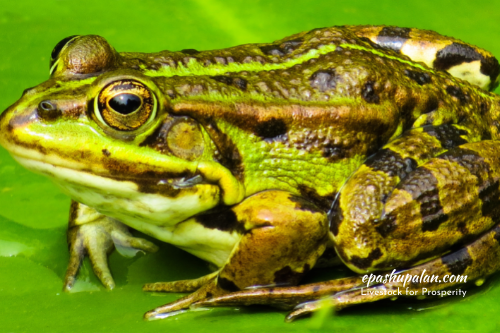
Clinical signs of toad poisoning are hypersalivation, vomiting and pawing at the mouth, cardiac irregularities such as cyanosis, weakness, pulmonary oedema, convulsions, prostration and collapse. In the case of B. marinus, death occurs within 15 minutes.
Treatment
There is no specific treatment but following steps can be taken as asymptomatic measures-
- Wash mouth with plenty of water.
- Give activated charcoal and osmotic purgatives.
- Administer atropine sulphate intravenously to check excessive salivation and bronchoconstriction.
- Administer cardioprotective agents such as propranolol (0.2 mg/kg).
- Administer antihistaminic, sedative or tranquilizers.
5. Snake Venom
Worldwide, venomous snakebites result in tens of thousands of deaths each year. Most fatalities occur in developing countries with high populations of both humans and venomous snakes and limited access to emergency medical care. Snake bite in animals generally occurs while grazing or hunting.
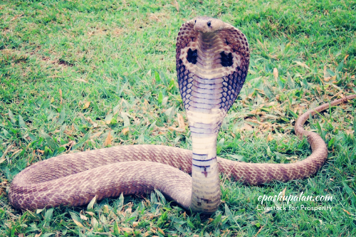
Snake venoms are complex mixtures of toxins- consisting of amino acids, polypeptides, glycopeptides and biogenic amines in addition to certain cations such as K+, Na+, Ca++, Mg++, Ni++, etc. Most active component of the venom is a peptide or polypeptide which may act enzymatically or non-enzymatically too. Generally, most of the snake venom produces two types of toxicity i.e., neurotoxicity and cardiotoxicity or haemotoxicity.
There are more than 3500 different species of snakes out of which more than 400 have been found to be poisonous and dangerous. Most of the poisonous snakes belong to the following 6 families-
Elapidae: Coral snakes, cobras, kraits
Crtotalidae: Rattle snake, pit vipers
Viperidae: The old-world vipers and adders
Hydrophidae: The true sea snake
Laticaudidae: Sea kraits
Colubridae: Boomslang, bird snake, etc.
Factors that affect toxicity of a snake bite include-
- Toxicity of the venom and quantity of venom injected
- Ratio of size of the animal and venom injected
- Species of snake
- Location of bite
- Species of animal involved. On the basis of body weight, horses are most susceptible
- Prompt availability of appropriate therapy
Venoms of snakes contain different fractions- necrotizing, anticoagulant, coagulant, neurotoxic, cardiotoxic and haemolytic fractions. The venoms of cobra and krait are mainly neurotoxic while that of vipers and rattle snakes are hemotoxic in nature.
Hemotoxins of pit vipers induce local tissue damage and injury to blood vessels, changes in blood cells, blood coagulation defects and shock. Cardiopulmonary dysfunction is also observed. There is hypotensive crisis. Local swelling at the site of bite is observed. Animals generally die without any previous history of illness.
Neurotoxin of cobra and krait induce paralysis of the cranial nerve of eyelids resulting into ptosis, paralysis of muscles of eyeball and respiration. Neurotoxins bind specifically to cholinergic receptors at the neuromuscular junction and produce curare like effects and respiratory paralysis. The affected animal shows salivation, hyperexcitability, mydriasis, asphyxia, gasping, recumbency, convulsions and death within 2-4 hours.
Assessment of severity of envenomation
| S.No. | envenomation | Signs |
| 1. | No envenomation | Absence of local or systemic reactions; fang marks (+/−) |
| 2. | Mild envenomation | Fang marks (+), moderate pain, minimal local oedema (0–15 cm), erythema (+), ecchymosis (+/−), no systemic reactions |
| 3. | Moderate envenomation | Fang marks (+), severe pain, moderate local oedema (15–30 cm), erythema and ecchymosis (+), systemic weakness, sweating, syncope, nausea, vomiting, anaemia, or thrombocytopenia |
| 4. | Severe envenomation | Fang marks (+), severe pain, severe local oedema (>30 cm), erythema and ecchymosis (+), hypotension, paraesthesia, coma, pulmonary oedema, respiratory failure |
Management of snake bite
- Keep the animal undisturbed.
- Apply a tight tourniquet above the site of bite to prevent further absorption and distribution of venom.
- Incise the local area of snake bite in the direction of the blood vessel and go for suction and infiltrate the area with 5% soap solution.
- Inject antivenin, antibiotics and antitoxins.
- If the snake has not been identified, administer polyvalent antivenin intravenously @ 10-20 ml/animal and also around the site of the bite.
- Supportive therapy against shock and cardiopulmonary disturbance should be given.
| The content of the articles are accurate and true to the best of the author’s knowledge. It is not meant to substitute for diagnosis, prognosis, treatment, prescription, or formal and individualized advice from a veterinary medical professional. Animals exhibiting signs and symptoms of distress should be seen by a veterinarian immediately. |


Be the first to comment