Introduction
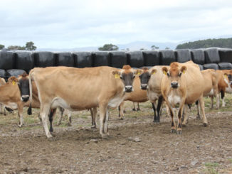
Brucellosis is one of the most important zoonotic diseases globally and continues to have a significant impact on animal as well as human health in world particularly in South America, Southern Europe, Africa, the Middle East and Asia (Pappas et al., 2006). Brucellosis is highly contagious zoonotic infection caused by the bacterial genus Brucella. The genus Brucella comprises 12 recognized species that include Brucella abortus (cattle), Brucella melitensis (sheep and goats), Brucella suis (pigs, hares, reindeer), Brucella canis (dogs), B. ovis (sheep), and B. neotomae (rodents) Brucella microti (voles), Brucella pinnipedialis (pinnipeds), Brucella ceti (cetaceans), Brucella papionis (baboons), Brucella vulpis (foxes), and Brucella inopinata (isolated from a human case—natural host unknown) (Whatmore et al 2012). These are small, Gram-negative, non-motile, non-spore-forming, aerobic, rod-shaped or cocco-bacilli in shape, present intracellularly in macrophages. They are shed in large numbers in the animal’s secretions and excretions like urine, milk, placental fluid, and other fluids. These organisms show tissue tropism towards the reproductive organs of host animals. It is one of the major organisms resulting in abortions and sterility in animals.
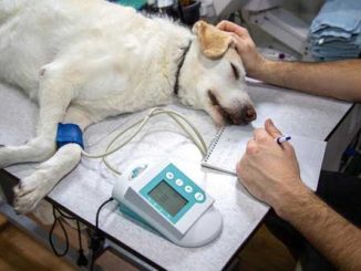
Canine Brucellosis is a highly contagious bacterial infection caused by the bacterium, Brucella canis (B. canis), a gram-negative cocco-bacilli that primarily causes reproductive failure in dogs. The disease spreads rapidly among closely confined dogs, and has been identified as the major reason for great economic loss in kennels (Gyuranecz et al 2011). Although dogs can become infected with Brucella abortus, B suis, or B melitensis on occasions, but these infections are sporadic in nature and occur mostly in population exposed to infected domestic livestock.

Transmission of Disease
Transmission occurs mainly via ingestion, inhalation, or contact with aborted fetuses or placenta, vaginal secretions, or semen (Carmichael et al 1988, Moore et al 1970). Like other Brucella species, B. canis also shows tropism for the reproductive tissue. Intermittent shedding of low concentrations of bacteria in reproductive fluids like seminal fluids and vaginal discharge is the primary source of infection in dogs (Hollett, 2006). Even after castration, dogs may still serve as a source of infection because the bacteria can persist in the prostate and lymphoid tissues.
In addition to these reproductive secretions, dogs can also shed the bacteria in the saliva, nasal secretions, and urine (Moore et al. 1969). Thus, genital, oronasal and conjunctival route are the major routes of transmission for this venereally transmitted disease in dogs. Minor routes of transmission include in utero, broken skin, blood transfusions, feces, milk, and fomites such as contaminated syringes, vaginoscopes, and artificial insemination equipment (Wanke, 2004 and Makloski , 2011).
Pathogenisis
The bacteria attaches to the mucous membranes, infilterate the epithelial barrier, and are taken up by the mononuclear phagocytic system, where they inhabit intracellularly in the macrophages. The virulence factor, primarily the type IV secretory system, is responsible for the penetration of the bacteria through the epithelial barrier and inhibiting the bactericidal myeloperoxidase-peroxide-halide system through the release of 5-guanosine and adenine (Chacón-Díaz et al 2015, Davidson et al 2014). Once the organism is present intracellularly, it travels through the reticulo-endothelial system to local lymph nodes (retropharyngeal, inguinal, superficial iliac), liver, spleen, and possibly bone marrow.
The bacteria then move into the blood stream causing intermittent bacteremia. The organism targets “steroid-dependent” reproductive tissues, including the prostate, testicles, epididymides, gravid uterus, and placenta (Davidson et al 2014). A mixed inflammatory response consisting of lymphocytes, plasmacytes, and histiocytes has been observed in these reproductive tissues (Brennan et al 2008). Focal coagulative necrosis of the chorionic villi, necrotizing arteritis, and numerous bacteria in trophoblastic epithelial cells can be found in the aborted placenta (Davidson et al 2014, Brennan et al 2008). Organisms can also spread to other non-reproductive tissues like the ocular tissue (eyes) causing anterior uveitis or endophthalmitis or the intervertebral disk causing discospondylitis in some cases (Chacón-Díaz et al 2015).
Clinical Signs
The clinical signs of B. canis infection are not pathognomonic. Dogs may be subclinically affected with no clinical signs other than loss of vigor. Occurrence of asymptomatic infections in dogs are not uncommon. During the early stages of brucellosis, enlarged lymph nodes are common, especially the submandibular and retropharyngeal lymph nodes. Although bacteremia may be persistent for several months, fever is not a typical clinical feature in the infected dogs (Santos et al 2021).
In male dogs, B. canis causes epididymitis, prostatitis, and orchitis. A dog with recently acquired infection will often have an enlarged scrotum or an enlarged testicle and may have a skin rash on the scrotum. As the disease progresses to chronic stage, the long term inflammation of testicles and epididymis can lead to unilateral or bilateral testicular atrophy, infertility, loss of libido and reluctance to mate. Sperm defects and head-to-head agglutination have been observed in the semen of dogs infected with B. canis. Oligospermia or azoospermia may also been observed in chronically infected dogs (George et al 1979).
B. canis can cause abortions and stillbirths in pregnant female dogs. Litters may contain both live and dead pups; however, live pups are often weak and frequently die soon after birth. Some congenitally infected animals appear normal, but may later develop brucellosis.
Mid- to late-term abortion (during days 45–59), followed by an odorless, brown-to-yellow, mucoid, sero-sanguinous vaginal discharge for 1–6 weeks is the typical clinical sign observed in bitches infected with B. canis (Carmichael et al 1968). Subcutaneous edema, hemorrhage, congestion and other nonspecific lesions can be observed in aborted pups. Pups that survive may be congenitally infected. These puppies appear healthy but are apparently bactremic and can shed the bacteria leading to dissemination of infection to other dogs and to humans also (Dentinger et al 2015). Also congenitally infected puppies that survive or dogs infected later in life may present arthritis, ocular disease, discospondylitis, urinary retention and osteomyelitis (Kornegay et al 1980).
Apart from abortion and still birth, embryonic death with resorption is also one of the important manifestations of brucellosis in bitches. This condition may go unnoticed and may appear as conception failure after a successful mating. Endometeritis has also been observed. It is possible for an infected bitch to abort and subsequently have normal pregnancies or intermittently experience reproductive failure; these dogs may serve as reservoirs for infection in B. canis–naive dogs. B. canis infection is the most common cause of reproductive failure in dogs, so it must be ruled out before investigating other causes of infertility or abortion (Carmichael et al 2012).
Diskospondylitis, an inflammatory disease of the spine characterized by infection of the intervertebral disc, adjacent vertebral endplates, and vertebral bodies, is another clinical condition associated with B. canis infection in dogs (Kornegay et al 1980, Henderson et al 1974). Infected dogs have a history of lameness, decreased physical activity, inappetance, spinal pain, neurologic dysfunction, muscle weakness, or any combination of these signs, caused by vertebral osteomyelitis and intervertebral disc infection (Anderson et al 1983). Incidence of diskospondylitis is higher in male than female dogs, perhaps because of a reservoir of bacteria in the prostate that results in intermittent bacteremia even in castrated males (Kerwin et al 1992, Hurov et al 1978). Occasionally, B. canis can also infect the eyes, kidneys, or brain. If the bacteria infects these other tissues, the signs will be related to the bodily system that is infected.
Post Mortem Lesions
Infected dogs may show generalised or localised lymphadenitis, splenomegaly and hepatomegaly. Scrotal edema, scrotal dermatitis, epididymitis, prostatitis, orchitis, and testicular atrophy and fibrosis may be detected in males, and metritis and vaginal discharge in females. Lesions related to localized infections, such as discospondylitis, osteomyelitis, meningitis, focal nonsuppurative encephalitis or abscesses in various internal organs may also be observed. Aborted fetuses are often partially autolyzed and may have evidence of a generalized bacterial infection, such as subcutaneous edema, congestion and hemorrhages in the abdominal region, bronchopneumonia, and degenerative lesions in the liver, spleen, kidneys and intestines. Some fetuses have no gross lesions.
Diagnosis
Diagnosis of Brucella canis infection is difficult due to the lack of highly specific and sensitive diagnostic assays. Humoral immunity induced by B. canis infection is poorly characterized, representing a challenge for the development of diagnostic tests (Bowden et al 1995).
The gold standard for diagnosis of B. canis infection is isolation of the causative agent (Wanke et al 2004). B. canis, as well as other Brucella spp., grows well-under aerobic conditions on conventional media, such as dextrose or tryptic soy agar under biosafety level 3 conditions due to its zoonotic potential (Wallach et al 2004). Farrell’s Medium is probably the most widely used selective medium for the bacteriological diagnosis of brucellosis (Farrell et al 1972). Samples of choice include Whole blood, aborted foetuses, semen and uterine or vaginal secretions. The best time for culturing Brucella is 2–4 weeks after infection, after demonstration of reproductive failure, when bacteremia is the highest.
Serologic tests evaluate antibody response against Brucella spp. cell wall antigens. Based on the structure of the O-polysaccharide subunit of lipopolysaccharide, Brucella spp. have 2 recognized appearances: smooth (considered more virulent; includes B. abortus, B. suis, and B. melitensis) and rough (B. canis and B. ovis). These differences in cell wall morphology form the basis of serological testing of the organisms (Rittig et al 2003).
Rapid slide agglutination tests (RSAT), tube agglutination tests (TAT) and immuno-fluorescent antibody tests (IFA) are used to rule out infection at early stages but have these tests have low sensitivity for chronically infected animals due to intermittent bacteremia (Keid et al 2009). These are followed by the screening tests like 2-mercaptoethanol rapid slide agglutination test and agar-gel immunodiffusion (AGID) (Mol et al. 2020).
Direct diagnosis can also be achieved by detecting B. canis genomic DNA in biological samples by polymerase chain reaction (PCR) (Bricker 2002, Keid et al 2007). Whole blood is the sample of choice for PCR. This technique is comparatively faster and not affected by the viability of bacteria or sample contamination. PCR used for diagnosis of canine brucellosis by targeting specific gene sequences that are conserved among Brucella spp. such as bcsp31, 16S ribosomal subunit and recA.
Several enzyme-linked immunosorbent assay (ELISA) protocols have been applied to the diagnosis of B. canis infection using antigens like B. canis surface antigens or cytoplasmic antigens (Lucero et al 2002). The complement fixation test (CFT) is considered a confirmatory test for B. ovis and B. abortus infection but has not been routinely used for diagnosis of canine brucellosis. Immunochromatographic assays have been developed for the diagnosis of B. canis infections. These are simple and rapid assays, but they have low sensitivity when compared to other traditional screening methods (Johnson et al 1992).
In cases with suspected discospondylitis due Brucellosis, Radiography or computed tomography is diagnostic in identifying end vertebral body osteomyelitis.
Treatment
First and foremost step is isolation of the dog tested positive for Canine brucellosis. Any animal that tests positive for Brucella must immediately be removed from the kennel or home in order to prevent the spread of the infection to other animals and humans. In case of multidog household or kennels testing of other dogs must be done immediately. Once the dog is has been isolated, it is important to destroy any and all material that may be infected with Brucella. Clean and disinfect the entire area to remove any persistent infection.
Long term treatment using a combination of two different antibiotics, such as tetracyclines or fluoroquinolones combined with aminoglycosides, can be used . But no treatment is completely effective at eliminating the bacteria, and any dog that has been infected with B. canis should be considered infected for life. Even when the organism seems to have disappeared, it may persist in tissues such as the lymph nodes, spleen, uterus and prostate. For this reason, euthanasia of infected animals is often recommended in some cases. Intact animals should be neutered, and some sources recommend that treated dogs be isolated even after treatment. Periodic serological monitoring might be able to detect rising antibody titer if organisms persist and begin to replicate again in treated animals. Therefore, the recommended course of treatment includes surgical sterilization, antibiotic treatment and strict isolation.
Also due to the zoonotic nature of the disease, it is important for people, who come in contact with breeding dogs, new born puppies, or aborted foetuses, to be cautious and practice good sanitation and disinfection of the area.
References
- Anderson GI, Binnington AG. Discospondylitis and orchitis associated with high Brucella titre in a dog. Can Vet J. 1983;24(8):249-252.
- Brennan SJ, Ngeleka M, Philibert HM, Forbes LB, Allen AL. Canine brucellosis in a Saskatchewan kennel. Can Vet J. 2008 Jul; 49(7):703-8
- Bricker B. PCR as a diagnostic tool for brucellosis. JVet Microbiol. 2002 Dec 20; 90(1-4):435-46.
- Carmichael LE, Joubert JC. Transmission of Brucella canisby contact exposure. Cornell Vet. 1988;78:63–73.
- Carmichael LE, Kenney RM. Canine abortion caused by Brucella canis. J Am Vet Med Assoc. 1968; 152:605–16.
- Carmichael LE. Canine brucellosis. In: Greene CE, editor. Infectious diseases of the dog and cat. 4th ed. London: Elsevier Health Sciences; 2012. p. 398–411.
- Chacón-Díaz C, Altamirano-Silva P, González-Espinoza G, Medina MC, Alfaro-Alarcón A, Bouza-Mora L, Jiménez-Rojas C, Wong M, Barquero-Calvo E, Rojas N, Guzmán-Verri C, Moreno E, Chaves-Olarte E, Brucella canis is an intracellular pathogen that induces a lower proinflammatory response than smooth zoonotic counterparts. Infect Immun. 2015 Dec; 83(12):4861-70.
- Davidson AP, Sykes JE. Canine brucellosis. In: Sykes JE, editor. Canine and Feline Infectious Diseases.1st ed. St. Louis, Missouri: Elsevier Saunders; 2014. pp. 512–519.
- Dentinger CM, Jacob K, Lee LV, Mendez HA, Chotikanatis K, McDonough PL, et al. Human Brucella canis infection and subsequent laboratory exposures associated with a puppy, New York City, 2012. Zoonoses Public Health. 2015;62:407–14.
- Farrell ID, Robertson L. A comparison of various selective media, including a new selective medium for the isolation of brucellae from milk. J Appl Bacteriol. 1972 Dec; 35(4):625-30.
- George LW, Duncan JR, Carmichael LE. Semen examination in dogs with canine brucellosis. Am J Vet Res. (1979) 40:1589–95.
- Gyuranecz M, Szeredi L, Rónai Z, Dénes B, Dencso L, Dán Á, et al. Detection of Brucella canis-induced reproductive diseases in a kennel.J Vet Diagn Invest. 2011;23:143–7.
- Henderson RA, Hoerlein BF, Kramer TT, Meyer ME. Discospondylitis in three dogs infected with Brucella canis. J Am Vet Med Assoc. 1974 Sep 1;165(5):451–455
- Hollett RB Canine brucellosis: outbreaks and compliance. 2006 Aug; 66(3):575-87.
- Hurov L, Troy G, Turnwald G. Diskospondylitis in the dog: 27 cases. J Am Vet Med Assoc. 1978;173:275–81.
- Johnson CA, Walker RD. Clinical signs and diagnosis of Brucella canisinfection. Compend Contin Educ Vet. (1992) 114:763–72.
- Keid LB, Soares RM, Vieira NR, Megid J, Salgado VR, Vasconcellos SA, da Costa M, Gregori F, Richtzenhain LJ. Diagnosis of canine brucellosis: comparison between serological and microbiological tests and a PCR based on primers to 16S-23S rDNA interspacer. Vet Res Commun. 2007 Nov; 31(8):951-65.
- Kerwin SC, Lewis DD, Hribernik TN, Partington B, Hosgood G, Eilts BE. Diskospondylitis associated with Brucella canis infection in dogs: 14 cases. J Am Vet Med Assoc. 1992;201:1253–7.
- Kornegay JN, Barber DL. Diskospondylitis in dogs. J Am Vet Med Assoc. 1980 Aug 15;177(4):337–341.
- Lucero NE, Escobar GI, Ayala SM, Lopez G.Sensitivity and specificity of an indirect enzyme-linked immunoassay for the diagnosis of Brucella canis infection in dogs. J Med Microbiol. 2002 Aug; 51(8):656-660.
- Makloski CL, Canine brucellosis management. Vet Clin North Am Small Anim Pract. 2011 Nov; 41(6):1209-19.
- Mol JPS, Guedes ACB, Eckstein C, Quintal APN, Souza TD, Mathias LA, Haddad JPA, Paixão TA, Santos RL. Diagnosis of canine brucellosis: comparison of various serologic tests and PCR.. J Vet Diagn Invest. 2020 Jan; 32(1):77-86.
- Moore JA, Gupta BN. Epizootiology, diagnosis, and control of Brucella canis.J Am Vet Med Assoc. 1970;156:1737–40.
- Moore JA. Brucella canisinfection in dogs. J Am Vet Med Assoc. 1969;155:2034–7.
- Pappas, G., Papadimitriou, P., Akritidis, N., Christou, L., and Tsianos, E. V. (2006). The new global map of human brucellosis. Lancet Infect. Dis. 6, 91–99.
- A. Bowden, A. Cloeckaert, M.S. Zygmunt, S. Bernard, L.A. Bowden, A. Cloeckaert, M.S. Zygmunt, S. Bernard. Surface exposure of outer membrane protein and lipopolysaccharide epitopes in Brucella species studied by enzyme-linked immunosorbent assay and flow cytometry. Surface Exposure of Outer Membrane Protein and Lipopolysaccharide Epitopes in Brucella Species, 63 (1995), pp. 3945-3952
- Rittig MG, Kaufmann A, Robins A, Shaw B, Sprenger H, Gemsa D, et al. Smooth and rough lipopolysaccharide phenotypes of Brucella induce different intracellular trafficking and cytokine/ chemokine release in human monocytes. J Leukoc Biol. 2003;74:1045–55.
- Santos, R. L., Souza, T. D., Mol, J. P., Eckstein, C., & Paíxão, T. A. (2021). Canine Brucellosis: An Update. Frontiers in veterinary science, 8, 121.
- Wallach JC, Giambartolomei GH, Baldi PC, Fossati CA. Human infection with M- strain of Brucella canis. Emerg Infect Dis. 2004 Jan; 10(1):146-8.
- Wanke MM, Canine brucellosis. Anim Reprod Sci. 2004 Jul; 82-83():195-207.
- Whatmore AM, Koylass MS, Muchowski J, Edwards-Smallbone J, Gopaul KK, Perrett LL. Extended multilocus sequence analysis to describe the global population structure of the genus Brucella: phylogeography and relationship to biovars.Front Microbiol. 2016;7:2049.
| The content of the articles are accurate and true to the best of the author’s knowledge. It is not meant to substitute for diagnosis, prognosis, treatment, prescription, or formal and individualized advice from a veterinary medical professional. Animals exhibiting signs and symptoms of distress should be seen by a veterinarian immediately. |


Be the first to comment