Introduction
Early and correct diagnosis is critical for prompt antifungal therapy implementation and reducing the usage of toxic antifungal drugs. Furthermore, having access to accurate and early diagnosis might minimize the use of empirical antifungal medication, lowering antifungal selection pressure and preventing the emergence of antifungal resistance. Standard approaches to the laboratory diagnosis of IFIs include direct microscopic examination in freshly obtained samples, histopathological demonstration of fungi within tissue sections, culture of the causative fungus and its further identification. However, these approaches are often not sufficiently sensitive and/or specific to diagnose Invasive fungal infections. So there is need for rapid methods which are sensitive and specific for diagnosis of fungal infections.
Diagnostic Methods for Fungal Infections
- Direct examination
- Fungal culture
- Radiology
- Non culture methods:
a) Serological methods- antigen detection, antibody detection
b) Tests for detection of metabolites
c) Tests for detection of CMI
d) Molecular methods
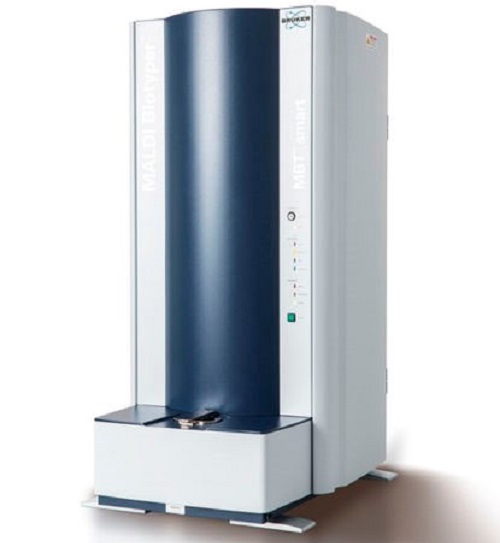
e) Others- MALDI-TOF MS
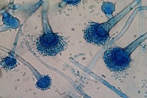
Direct Microscopic Examination of Clinical Specimen
Microscopy techniques include fresh and stained examination of microbiological samples like KOH, India ink etc. Before the growth of fungal cells in culture, a preliminary diagnosis may be made, and suitable antifungal drugs can be started. Observing fungal cells in a clinical specimen rather than isolating in a culture may be more useful as a diagnostic criteria. These diagnostic techniques have significant drawbacks, one of which is their poor sensitivity, which means that the fungal element is only detected when it is highly numerous in the sample to be examined, i.e. when the infection has advanced. It is also impossible to determine the species.
Calcoflour White can be added with KOH, which binds specifically to β 1-3 D-glucan and gives brilliant apple-green fluorescence when exposed to the shorter wavelengths of ultraviolet light. India ink is a negative staining technique, useful for indicating the presence or absence of extra cellular polysaccharide capsule of fungal cells. Histopathological sections stained with haematoxylin and eosin, periodic acid– Schiff and silver stains, may help in detecting the presence of fungal element in many cases of deep mycosis.
Gormori’s Methenamine Silver Stain: It is based on the liberation of aldehyde groups and their subsequent identification by the reduced silver method. It is used for demonstration of polysaccharide content on the fungus in tissue sections.
Microbiological Culture
Blood agar, Sabouraud dextrose agar with or without cycloheximide, malt extract agar, cornmeal agar, and brain heart infusion agar are some of the most widely used culture media for fungal growth. At 25°C and 37°C, the cultures are incubated. Selective media, such as Sabouraud dextrose agar containing antibiotics like chloramphenicol and gentamicin, are indicated for samples with a polymicrobial infection or commensal bacterial flora.
Characterization to species level is essential for all clinical isolates collected from deep tissue sites and can be done using conventional methods of classification based on morphological features like chlamydospore formation, germ tube formation, adhesive tape preparation and slide culture and biochemical features like sugar fermentation, sugar assimilation and urease test.
Radiology
High-resolution CT and Magnetic Resonance Imaging (MRI) scans make it possible to detect both pulmonary and extrapulmonary invasive aspergillosis and other invasive fungal infections.
Non-Culture-Based Diagnostic Methods
Serological Diagnosis
Antibody detection is helpful in determining the prognosis of fungal disease and therapy response. However, because of the significant cross reactions caused by the complex structure of antigens, specificity and sensitivity are poor. Additionally, antibodies may be present in the absence of infection due to past exposure to the fungus as a saprophyte or as a commensal. As a result, antigen detection is a viable option for resolving these problems. Various serological techniques used are
- Latex particle agglutination
- Immuno-diffusion
- Countercurrent immune-electrophoresis,
- Immuno-fluorescent antibody
- ELISA
- Western blotting
Test for Detection of Metabolites
This is to establish the presence of a distinct fungus metabolic byproduct in patients’ bodily fluids. It can be utilised as a prognostic indication as well as a serum marker for Candida spp. Gas chromatography (GC) in serum and/or urine can be used to identify D-arabinitol production, or D-arabinitol can be quantified enzymatically and detected spectrophotometrically.
Tests for detecting CMI
Skin testing can be used to establish etiological diagnosis, especially in nonendemic areas, conduct epidemiological surveys, and serve as a prognostic indication, as a positive skin test can become negative with severe or disseminated diseases. Aspergillus fumigates, Histoplasma capsulatum, Sporothrix schenkii, and other fungi are tested on the skin. However, in endemic areas and owing to cross reactivity caused by antigen sharing, it might provide false positive results. Due to the early stage of infection and a faulty CMI, false negatives might arise.
Molecular methods
Molecular identification, based on the sequencing of several DNA targets or fragments, has shown that classification by conventional techniques is not reliable for some moulds, especially the less common species. Detection and identification of fungal pathogens by DNA-based methods can yield results sooner than cultivation and also the sensitivity and specificity are higher than cultures and serologic tests for diagnosing infections. Also DNA results have correlated with clinical improvement and effect of treatment, in patients with invasive aspergillosis, invasive candidiasis, and cryptococcal meningitis. Furthermore, this strategy made it possible to decrease the administration of antifungal drugs by reducing their empirical use.
However there are certain disadvantages associated with molecular techniques:
- Molecular diagnostic methods may not distinguish colonization from infection.
- False-positive results of 31% and ranging from 8 to 38% have been reported in the diagnosis of candidiasis and aspergillosis due to colonization of the patients’ airway by conidia and/or hyphae and the contamination of sample and/or reaction buffers by environmental fungi.
- False-negative results of 21% of patients with positive blood cultures for Candida species, due to the low sensitivity of detection of single-copy gene, hence it is necessary for these techniques to incorporate an internal control.
In the last decade, effective amplification platforms, probe development and various quantitative PCR technologies have revolutionised research on fungal detection. Some of the latest techniques employed in the detection of fungi are:
• Fluorescence in situ hybridisation (FISH)
• DNA array technology
• Multiplex tandem PCR
• Real time PCR
• PCR- ELISA
• Random Amplified Polymorphic DNA (RAPD)
• Loop-Mediated Isothermal Amplification (LAMP)
Other Techniques
MALDI-TOF MS
The methodology of identification of yeasts and filamentous fungi by MALDI-TOF MS is still evolving. Most species of Candida and filamentous fungi such as Aspergillus species can be identified rapidly and accurately by this technique.

Be the first to comment