Introduction
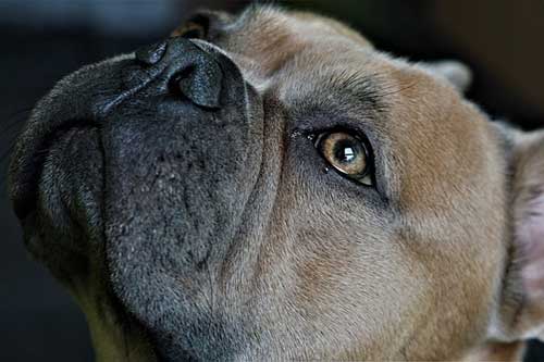
Epiphora or tearing is the presence of watering eye or ocular (serous) discharge. The etiology of epiphora is divided into two categories. They are (i) increased (reflex) tearing and (ii) impaired tear drainage/reduced tear outflow.
Reflex tearing is usually due to pain and inflammation from corneal ulcer, glaucoma, uveitis and secondary to lens displacement and foreign body whereas primary hypersecretion of the lacrimal glands is rare (Plummer, 2013).
Reduced tear outflow or impaired tear drainage are due to congenital or acquired causes. Congenital causes for reduced tear outflow or impaired tear drainage are atresia or absence of or abnormal displacement of the punctum and poor conformation of tight ligament at medial canthus results in tear pump dysfunction caused by eyelid laxity or medial entropion and or presence of hairy caruncles in case of Toy Poodle breed dogs. Acquired causes for reduced tear outflow or impaired tear drainage are dacryocystitis / dacryorhinocystitis, Dacryocystitis / dacryorhinocystitis is the inflammation of lacrimal sac and nasolacrimal duct usually results from presence of foreign body. Other causes are nasolacrimal duct obstructions (NLDO) – obstruction at any portion of the nasolacrimal drainage system. NLDO may be congenital or acquired. FHV-1 (Feline Herpes virus) is the most common cause of adhesions and nasolacrimal obstruction in cats. Other causes for NLDO are subsequent to eye injury, tumors etc., Epiphora due to combination of causes are also not uncommon.
Diagnostic approach
- Ocular examination: complete and thorough ocular examination to rule out causes of reflex tearing.
- Evaluation of nasolacrimal system for reduced drainage
Schirmer Tear Test (STT)
- STT is the preliminary test used to assess the aqueous layer of the tear film only. If STT is more than 25 mm/min, check for blepharospasm presence. Stimulated lacrimation could be due to blepharospasm resulting from pain/irritation. Distichiasis, ectopic cilia, allergic conjunctivitis, blepharitis, eyelid masses, keratitis, corneal ulcer, anterior uveitis, acute glaucoma, anterior lens luxation, kerato-conjunctivitis sicca (KCS) and medial canthal trichiasis. These are primary causes for blepharospasm induced lacrimation.
- When the STT is within the normal range, more likely to be impaired drainage/reduced tear outflow.
- Check for the presence and patency of lacrimal puncta and examine for foreign body
- When the lacrimal puncta is present with patency, Jones test/fluorescein dye passage test is performed to assess the nasolacrimal duct patency and irrigation of nasolacrimal duct. Fluorescein applied to eye should appear at nasal mucosa within 30 seconds to 5 minutes.
- Congenital absence of puncta, imperforate puncta, micropunta, fibrosis from chronic inflammation or traumatic laceration resulting in stenosis or occlusion, neoplasia of surrounding tissue resulting in invasion or occlusion, cyst and symblepharon resulting in blockage of lacrimal puncta.
- Punctal catheterization and flushing – normograde (from the eye down to the nose) in dogs and cats
- Procedure: use topical anaesthetic 0.5% proparacaine, 22-24 G IV catheter stylet is removed and a 3 ml syringe with saline. Introduce catheter into puncta occlude other puncta and flush gently.
Reference
- Plummer, C. E. Proptosis reduction. Clinician’s Brief. September 2013. Page no. 67-71.

1 Trackback / Pingback