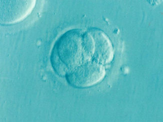
Embryo searching
- Filter to rinse with 20 ml fluid using syringe and needle
- Bottom dish (below the filter) use to collect the fluid and for embryo searching
Handling abnormal cervix
- ‘Y’ shaped cervix
Single vaginal opening , however, two opening in the body
Such condition horn flushing and horn insemination advised
- Inverted ‘Y’ Shaped cervix
Two vaginal opening with single uterine opening – Do not hamper flushing or AI
- Double barrel cervix
Two separate vaginal and uterine opening – Separate flushing of both horns to be followed
Damages
- Brown blood clot in the first flush
Indicate trauma to uterine body by AI gun. Brown colour indicate old injury
May also reflect that oocytes were not fertilized
- Disappearance of infuse fluid
Injury by AI gun that do not heal (brown clot)
Fresh rupture by catheter over inflation (red clot)
- Leaky valve (UTJ)
Happens when many follicles remains unovulated (high E2)
More E2, leads to improper closer of UTJ
Fluid leaked from oviduct to abdomen
- Catheter patency
Squeezing the bifurcation area gently 2-3 times
To and fro movement of bubbles at the outflow end of the tubing reflect patency of catheter
Reposition catheter if no patent
- Catheter washing when mucous is trapped in the catheter
While passing through difficult cervix mucous get trapped in the catheter
Mucous is enemy to embryo
Cuff bifurcation area with thumb and four fingers (blocking)
Allow 15 ml fluid to enter the catheter and Flush out and discard
Selection of recipient for transfer
- Size
Should be with the expected calf size to avoid dystocia and Heifer should be over 350 kg
- Milk–Should have sound udder
- Prior history – Regular calving
- Lactation
Good body condition lactating animals (major source of recipient)havig 40-50 days post partum
Dry cows are excellent recipient, however avoid problematic animals& already weaned calf
- BCS – Not too fatty , ideally BCS 3 to 3.5
- Mineral deficiency – As per the area deficiency, incorporate area specific minerals
- Low stress
Adequate shelter in cold and heat and Do not allow to go them in mud
Bio security
Should be disease free
No such disease as BVD, Neospora canis, JD, Bovine leukosis
- Vaccination – Against BVD, IBR, Parainfluenza 3, Leptospira
Recipient synchronization
- Natural or induced heat do not have any impact on pregnancy • Fixed time ET (FTET) also work good, however, have low pregnancy rate but yield is more
- Explanation :
- If 100 animals synchronized and observed for heat• 85% will be observed in heat
- 75% will have good CL & will be selected for transfer
- If pregnancy rate is 60% the calf obtained will be 45
- For timed ET
- Out of 100 animals, good CL will be 90%
- If pregnancy rate considered as 55% than calf obtained will be 49.5
Acceptable synchrony of recipient
- Fresh embryo tolerate greater degree of asynchrony than frozen
- 24 h before to 48 h after donor heat , recipients have been used
- Day 7 recipients are always best
- For frozen embryo – 5.5 – 7.0 day heat used
- For fresh emrbyo – 5-8 days heat used
- Grade 1 embryos used for cryopreservation
- Grade 2 & 3 embryos used for transfer but Day 8 embryos are to be avoided
Recipient preparation
- Should be full rumen and not fasting
- Recipient in chute
- Epidural -3- 5 ml lignocaine
- Palpate ovary quickly
- Transfer embryos if good CL and normal uterus palpated
- Transfer should be in horn ipsilateral to ovary having CL
- Provide comfortable environment to recipient
- Transport to short distance do not cause any harm
- Site of transfer – Cranial 1/3rd of the horn (deep transfer)
Avoid any trauma during transfer, as it will lead to PG release
- Recipient restraining – Properly squeezed with Minimum movement
- Footing – Firm footing in chute (no metallic floor)
- Climate – Comfortable and cool but Avoid air blown through fan or plenty of air flow
Cooler part of the day should be used for transfer
- Embryo deposition
Time spent in passing the gun through cervix do not effect pregnancy rate
Time spent in passing through horn adversely affect pregnancy rate
Double sheath gun is used for transfer
- Frozen thawed ET
Transfer similar as fresh embryo transfer, however, frozen-thaw are transfer immediately while fresh can be retain for 6-8 h
- Thawing – Water thaw – Air thaw – Combination (water + air)
- Water thaw : straw directly transfer from LN2 into water bath or thawing unit
Chances of zona cracking
- Air thaw: straw after taking out from LN2 , kept in air till completely thawed
- Combination: after taking out from LN2, straw kept in air for 5-6 seconds and than transferred to water bath – Prevent zona cracking
Cracked zona embryo can not be used for export
Some time crack zona is helpful in easy hatching
For cryopreserved embryos, combination therapy is commonly used
- ET gun – IMV 52 cm ET gun with blue sheath
- Success rate
5% difference with fresh embryo (AETS: American Embryo Transfer Association)
Evaluation of embryos
- Morphological evaluation performed for three reasons
To differentiate between embryo & unfertilized ova (UFO)
Determining the developmental stage coinciding with the expected development
To unable technician to have sufficient information on which to base the decision to transfer or cryopreserve
Parameters considered for embryo evaluation
- Embryo size & shape • Presence of extruded / degenerated cells
- Colour characteristics, number and compactness of the cells (blastomeres)
- Integrity of zona pellucida • Presence or absence of vacuoles in cytoplasm of blastomeres
Grading and classification (IETS)
- IETS established in 1974 (Denver, Colorado, USA)
- Two digit coding system to describe embryo & their characteristics – 1st digit represent embryo stage – 2nd digit represent embryo quality – Both digit separated by hyphen ‘ – ’
- Embryo stage code ranges from 1- 9 (unfertilzed to expanded hatched blastocyst)
- Embryo quality code ranges from 1- 4 (excellent /good to dead/degenerated)
Microscope for embryo evaluation
- Good microscope having high resolution to be used
- Steriomicroscope with maximum magnification of 50x should be employed
- Embryos initially searched in 10-15x • For zona & cytoplasmic evaluation – 50x
- Microscope should have long working distance (distance b/w objective to stage)
- Stage plate should be clear (No frosted plate) • No clamp or clip on the stage (free stage)
- Illumination from beneath the stage • Bright field illumination as standard but should also have facility for dark field illumination (helpful when searching through cloudy fluid)
Normal development of embryo (in-vivo)
- Embryo diameter – 150-190 μm • Zona thickness – 12-15 μm
- The size remain unchanged till blastocyst stage.
- Zona function – Provide shape to embryo – Provide receptor for sperm
Block accessory sperm (cortical reaction) – Rupture of zona leads to hatching
Zona is one of the landmark in recognizing the embryos
Stage of embryos encountered on day 7 :
Morula – blastocyst • Individual cells of embryos called blastomeres
- Morula – Blastomeres appears like cluster of grapes & individual blastomere could not be differentiated
- Compact Morula – Cells differentiated as ‘inside’ & ‘outside’ part of embryos
- Blastocyst
Two types of cells differentiated
Outer ring of trophoblast cells form the outermost layer of placenta
Inner clump of cells called inner cell mass (ICM) form entire foetus as well as most of the layer of Placenta
Posses cavity called blastocoele\
Stages of embryonic development recovered on D-7
- Stage code 1 : 1 cell
Getting a single cell between D 6-8 indicate unfertilized ovum (UFO)
Stage may confused with compact morula – All UFO many not have similar morphology
- Normal UFO
Perfectly spherical zona – Spherical vitelline membrane
Normal granular cytoplasm – Moderate pervitelline space
- Other UFO
Shows signs of degeneration (variable degree) – Cytoplasmic fragmentation
May give illusion of blastocoele or cell division
Extreme condensation (resemble compact morula)
- Stage code 2: 2-cells to 12-cells
Any embryo representing these cells number between day 6 –8 is not coordinating with expected development and considered dead or degenerated
Developmental delay, not fit for transfer or cryopreservation
- Stage code 3 : Early morula (latin – mulbery)
Minimum 16 cells to considered morula – Some blastomere visible some not
- Stage code 4 : compact morula
Blastomere coalesed to form a compact-light ball of cells-Individual blastomere not visible
Cells can be allocated as ‘inside’ or ‘outside’ part of emrbyos
Occupy 60-70% of perivitelline space.
- Stage code 5 : early blastocyst – Fluid filled cavity appears
Cells start differentiating between zona & blastocoel (but no clear ICM)
No change in thickness of zona – Cells occupy 70-80% of perivitelline space
- Stage code 6 : blastocyst
Cells clearly differentiated – Trophoblast layer, ICM, Blastocoele
Zona thickness unchanged
No perivitelline space (except where cells are partially collapsed)
- Stage code 7: expanded blastocyst
Increase in overall diameter ( 1.2-1.5 times of original 150-190 µm)
Zona pellucida thining 1/3rd of original (12-15 µm)
No perivitelline space– Expanded blastocoele stage embryo frequently appears collapsed
- Stage code 8 : hatched blastocyst
Embryo can be seen undergoing hatching process or hatched
Embryo has blastocel cavity or may be collapsed
Difficult in identification of this stage as may confuse with endometrial debris
- Stage code 9 : expanded hatched blastocyst
Similar to stage 8, however has larger diameter (size) – Difficult to get this stage on D 7
Embryo quality grade
- By visual estimation of embryo morphological characteristics
- Assessment features
Uniformity of blastomeres (size, shape & color) – Presence of extruded cells
Presence of dead/degenerated cells – Degree of cytoplasmic granulation
Presence of cytoplasmic vacoule – Presence of cytoplasmic fragmentation – Zona integrity
- Code 1 : excellent /good
Symmetrical and spherical embryo mass
Individual blastomere uniform in size, color , density
Developmental stage of embryo coinciding with the expected development age.
85% normal blastomere – Zona spherical and smooth (no concave / flat surface)
- Code 2 : Fair
Moderate irregularities – 50% blastomere normal and intact
Mild unevenness in cytoplasmic pigmentation with in individual blastomeres
- Code 3: Poor
Major irregularities in shape, size & color of individual blastomeres (25% cells intact)
Extreme unevenness of cytoplasmic pigmentation – Presence of vacuoles in the blastomeres
- Code 4: Dead or Degenerated
Extreme dark cytoplasm– If no embryo at this stage of collection reach to morula stage than embryo should be considered as dead/degenerated
Embryo Evaluation
- Recovered embryo transfer from filter to search Petridis
- Embryo searched in flushing / recovered media at 10 -15 x (steriomicroscope )
- Searched embryos transfer to holding media in another Petridis Evaluation for morphology at 50 x
- Commonly recovered embryo stage on day 7 : compact morula, early blstocyst & blastocyst
- Embryo recognized by zona (translucency & refract light)
- Two digit code : first representing stage than quality
- Example : 6 – 1 code means, blastocyst stage –excellent /good quality
Note : Why there is difference in embryo stage and quality : time of ovulation differ
Non-transferable embryos • Key identification features
Normal embr U
| Normal Embryo | UFO |
| Vitelline membrane smooth & spherical | Membrane fragmented /degenerated |
| Cellular cytoplasm | Granular cytoplas |
| No perivitelline space | Moderate perivitelline space |
- Degenerated ova appears similar to compact morulla
- Fragmentation: referred as cytoplasmic blebbing is a process during which some of
cytoplasm (which does not contain any chromosome or chromosomal DNA) segregate itself from ovum, resulting in a specimen that appears multicellular
- Can be checked by DNA staining • Resemble 2-4 cells stage embryos
- Getting a 2-4 cells stage embryo at this time period is illogical.
Dead / Degenerated embryos
Any embryo between 2 cell to 12 cells stage collected on day 7 is considered as dead /degenerated. – Indicate slow and retarded development which will not leads to pregnancy
Embryos whose blastomeres are not fused to one another (lack of tight junction formation) is also considered as dead /degenerated
Transferable embryos
- Embryos despite having normal quality may have many deviations. These may includes
Irregular size of blastomeres – Large/ multiple vacuoles within the cytoplasm of blastomere
Degeneration of one or more blastomere
Extruded blastomeres (some cells not forming tight junction)
Damaged or mishapen zona pellucida
- Extruded cells
Adhered blastomere communicate with one another and participate in development of embryos
Non adhered cells considered as extruded
Size and number of extruded cells directly impact embryo grade and pregnancy rate
Extruded cells do no divide
Extruded cells pressed with zona layer (stage 6-7) difficult to identify
Rolling of embryo assist in identification
- Misshapen or flat zona pellucida
If zona is not spherical than considered as misshapen having flat or concave surface
Such zona interfere with rolling & allow embryos to stick
The defect is sufficient to lower the embryo 1 grade than what actually observed
- Lipid in cytoplasm
Abnormal accumulation of lipid droplet (vacuoles) in the cells is not good
Presence of more than few vacuoles in the cells cytoplasm is grade lower in quality
- Cracked zona pellucida
Cracking usually observed at the time of hatching
Cracking of zona may occur at early stage
Cracked zona embryo can not be washed properly
Embryos not fit for export, however can be frozen well
- Irregular shape of cells comprising the embryos
Commonly observed in embryos of blastocyst stage
Formation of blastocoele cavity leads to irregularity
Collapsing of blastocoele is considered normal physiology.
Not a cause for lowering the grade
- Materials adhering to zona pellucida
Some embryos may have adhered material on the outer surface of zona
- Adhered material may include – Mucus – Individual cells – Clumps of tissue
- Adhered material also reflects signs of endometritis in donor
- Cumulus cells / endometrial cells constitutes the adherent materials (do not separate during transport)
- Adherent materials can be washed successfully or even can be washed with 0.25% solution of trypsin • The quality of embryo is not affected by adherent materials , however, such embryos are not fit for commercial use or export
Challenges to accurate embryo grading & classification
- UFO with compact morula
Good microscope – Higher magnification help in accurate grading
- UFO with blastocyst
Happens when a portion of UFO degenerate and become lighter resembling blastocoel
Check trophoblastic layer surrounding the blastocoele – ICM cells in blastocoele
All are absent in UFO
Embryo evaluation tips
- Most common disagreement amongst workers are between grades (excellent /good to fair to poor quality) rather than stage
- Rolling of embryo will help in evaluation of zona & embryo make up
- Each embryos carefully examined with zoom in and out way and rolled (to observe all sides as it’s a two dimensional figure)
- Effectiveness of morphological embryo evaluation
Photograph helps as a learning tool, however, actual evaluation needed
Video images further assist in the process
Reports based on video images showed agreement by experienced technician for
- Embryo stage – 89% • Embryo quality – 68.5% (Farin et al., 1995, Theriogenology
- The difference in agreement is because of lack of rolling as can be done in a petridish
- Success to embryo evaluation depends upon
Appropriate training (have seen all stage & grade of embryos)
Ample experience – Proper equipments (good microscope)
- Embryo stage has little influence on pregnancy rate when transfer between compact morulla to expanded blastocyst for in-vivo fresh embryo
- Similarly stage code 4,5 & 6 of in-vivo derived embryo survived freezing – thawing process well , however, grading is important


Be the first to comment