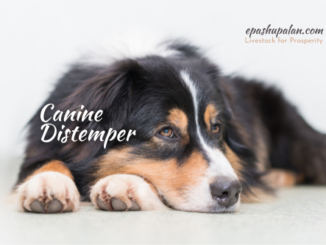
Abstract
The members of the genus Microsporum is primarily considered a zoophilic dermatophyte, mainly M. canis. One of the most prevalent superficial fungal diseases in dogs and cats is Microsporum infection. The most frequent method of transmission involves direct contact with infected human and animals. The common clinical symptoms include multifocal alopecia, mild to severe pruritus, and round, scaly lesions with erythematous and scaly borders. Clinical history, physical examination, and diagnostic procedures like Wood’s lamp, direct microscopic examination of infected hairs and/or crusts, fungal culture, and biopsy are used to diagnose Microsporum infection in pet animals. A combination of topical, systemic, and environmental disinfection is needed for the effective clinical and mycological cure of Microsporum infection
Keyword: Dermatophyte, Microsporum, Ringworm, Wood’s lamp
Introduction
Small animal dermatophytosis is a major concern for pet breeders and owners with Microsporum being the most common dermatophyte affecting them. Contact with contaminated objects, such as furniture or grooming tools, or with infected animals can spread Microsporum mediated infections either directly or indirectly. The infective propagules (spores) of Microsporum spp. infect the stratum corneum and hair after adhering to the epidermis of a susceptible host. Young stressed, or elderly animals are more likely to be infected. (DeTar LG et al., 2019). Microsporum canis is the primary cause of feline infections, and with the right care, the majority of infected cats make a mycological recovery within three weeks (Moriello KA et al., 2020).
The prevalence of Microsporum infection in dogs and cats ranges from 4 to 10% and more than 90%, respectively, but this may vary depending on regional variations and other epidemiological factors (Cabanes FJ et al., 2000; Frymus et al.,2013). Young dogs, those between the ages of 6 and 18 months, are more prone to infection than those between the ages of one and a half and three years (Singathia R et al., 2014). Male dogs are more susceptible to contracting the disease than female dogs (Singathia et al. 2014 and Bhardwaj et al. 2012). Infection rates in Yorkshire terriers were significantly higher than those in other breeds, especially those caused by M. canis, indicating breed predispositions to canine dermatophytosis (Bhardwaj et al. 2012). Seasonal variations may have an impact on the frequency of a particular agent in cases of small animal dermatophytosis. In contrast to N. gypsea, which is more common in the spring and summer, M. canis ringworm is more common in the fall and winter (Lewis DT et al., 1991).
Clinical signs
Common clinical symptoms include multifocal alopecia, mild to severe pruritus, and round, scaly lesions with erythematous and scaly borders.

Diagnosis
Clinical history, physical examination, and diagnostic procedures like Wood’s lamp, direct microscopic examination of infected hairs and/or crusts, fungal culture, and biopsy are used to diagnose Microsporum infection (Gross TL et al., 1992; Moriello KA et al., 2019).
Treatment
Microsporum infection in dogs and cats requires a combination of topical, systemic, and environmental disinfection to be successfully treated.
- Topical Treatments consists of hair clipping, dips, shampoos, and rinses
- Systemic treatments with azole or allylamine drugs
The best drugs for systemic therapy are those that accumulate in skin and keratin and are keratinophilic and lipophilic. Currently, the most efficient systemic treatments for both canine and feline dermatophytosis are oral itraconazole or oral terbinafine (Moriello KA et al., 2017). Ketoconazole, miconazole, and griseofulvin are also used in fungal infection of canine and felines.
- Environmental decontamination
The recurrence of Microsporum infection can be lessened with weekly cleaning and topical therapy. Studies have shown that weekly cleaning is very effective in removing infective spores from contaminated environment. The hardest part of the decontamination procedure is the actual cleaning, which entails the removal of dirt and hair. Cleaning can be done using commonly available household detergents. A 1:100 solution of household bleach or accelerated hydrogen peroxide can be used to disinfect hard surfaces. To maximise spore removal, soft fabrics should be machine-washed on the longest cycle possible (Moriello KA et al., 2013,2015, 2019).
Conclusion
Although Microsporum spp. mediated infections are curable with antifungal drugs and hygiene, early diagnosis should be done based on clinical signs and fungal culture to avoid the progression of the disease and transmission to susceptible animals. Wood’s lamp examination can be used for screening Microsporum canis infected animals. A combination of topical and systemic treatments are necessary for the majority of affected patients. The best oral treatments are terbinafine and itraconazole, which can be combined with miconazole and chlorhexidine-containing shampoos, topical lime-sulphur dips applied twice a week, and other treatments. It is best to use a combination of clinical sign resolution and a negative culture to determine whether a patient has fully recovered.
References
- Bhardwaj RK, Taku AK, Ahmad I. 2012. Therapeutic management of dermatophytosis in canine. Indian Vet. J. 89: 61-62.
- Cabañes FJ. 2000. Dermatophytosis animales. Recientes avances. Rev. Iberoam Micol. 17: S8–S12.
- DeTar LG, Dubrovsky V, Scarlett JM. 2019. Descriptive epidemiology and test characteristics of cats diagnosed with Microsporum canis dermatophytosis in a northwestern US animal shelter. J Feline Med Surg. 21(12): 1198–1205.
- Frymus, T., Gruffydd-Jones, T., Pennisi, M.G., Addie, D., Belak, S., Boucraut-Baralon, C., Egberink, H., Hartmann, K., Hosie, M.J., Lloret, A., Lutz, H., Marsilio, F., Mostl, K., Radford, A.D., Thiry, E., Truyen, U. and Hornizek, M.C.D. 2013. Dermatophytosis in cats: ABDC guidelines on prevention and management. J. Feline Med. Surg., 15(7): 598-604.
- Gross TL, Ihrke PJ, Walder EJ. 1992. Veterinary Dermatopathology: A macroscopic and microscopic evaluation of canine and feline skin disease. St. Louis: Mosby – Yearbook. 241-246.
- Lewis DT, Foil CS, Hosgood G. 1991. Epidemiology, and clinical features of dermatophytosis in dogs and cats at Louisiana State University: 1981–1990. Vet. Dermatol. 2: 53–58.
- Moriello KA, Coyner K, Paterson S, Mignon B. 2017. Diagnosis, and treatment of dermatophytosis in dogs and cats: Clinical Consensus Guidelines of the World Association for Veterinary Dermatology. Vet Dermatol. 28(3): 266–268.
- Moriello KA, Kunder D, Hondzo H. 2013. Efficacy of eight commercial disinfectants against Microsporum canis and Trichophyton spp. infective spores on an experimentally contaminated textile surface. Vet Dermatol. 24(6): 621–623, e151–152.
- Moriello KA, Stuntebeck R, Mullen L. 2020. Trichophyton species and Microsporum gypseum infection and fomite carriage in cats from three animal shelters: a retrospective case series. J Feline Med Surg. 22(4): 391–394.
- Moriello KA. 2019. Decontamination of 70 foster family homes exposed to Microsporum canis infected cats: a retrospective study. Vet Dermatol. 30(2): 178–e55.
- Moriello KA. 2020. Immediate and residual antifungal activity of compounds used for whole body and adjuvant topical therapy against Microsporum canis: an in vitro study. Vet Dermatol. 31(4): 272–e64.
- Singathia R, Gupta S, Yadav R, Gupta Y, Lakhotla RL. 2014. Prevalence of canine dermatophytosis in semi-arid Jaipur. Haryana Vet. 53(1): 43-45.
| The content of the articles are accurate and true to the best of the author’s knowledge. It is not meant to substitute for diagnosis, prognosis, treatment, prescription, or formal and individualized advice from a veterinary medical professional. Animals exhibiting signs and symptoms of distress should be seen by a veterinarian immediately. |


Be the first to comment