Inflammation of one or more quarters in the udder or inflammation of parenchyma of mammary gland due to multiple etiological agents. Mastitis is the one of the economical important disease of cattle and buffalo caused by various organisms. The disease has worldwide distribution. High milk yielding animals with poor management is one of the important risk factor for mastitis. It is multi-etiological complex disease. Mastitis is second most important disease for culling of the female animals after the reproductive problems. It is impossible to eradicate the disease, but with good managemental practices the disease occurrence can be reduced. It is the most costly disease in dairy industry. According to Kennedy and Miller (1993), mastitis is expressed by tissue injury caused by tissue invasive or toxigenic organisms, which become dominant due to upset of balance in microbial population. Incidence of clinical mastitis is about 5-10%. 70% of mastitis is caused by bacteria, 2% caused by yeast and moulds and 28% caused due to physical trauma and related factors.
Predisposing factors for mastitis
- Poor hygiene in the farm.
- Trauma to teat and/or udder.
- Low immunity in cow.
- Viral disease infection like Foot and mouth disease, Cowpox, Lumpy skin diseases predisposes animal for mastitis.
- Poor plane of nutrition.
- Faulty milking practices.
- Defective milking machine.
- Injury caused due to bite caused by calf.
- Early stage of lactation.
- Mastitis highest in summer.
- Deficiency of vitamins A and E, Cu, Zinc, Selenium.
- Extreme environmental conditions.
- Dirty bedding materials.
- Wet floor in the shed.
Causes of mastitis
- Staphylococcus aureus,
- Streptococcus agalactiae
- Mycoplasma bovis
- Corynebacterium bovis
- Coagulase-negative Staphylococci
- Escherichia coli
- Klebsiella
- Enterobacter spp
- Mycobacterium tuberculosis
- Arcanobacterium pyogenes
- Streptococcus uberis
- Streptococcus dysgalactiae
- Streptococcus equinus
- Nocardia spp,
- Pasteurella species.
- Mycobacterium bovis,
- Bacillus cereus
- Pseudomonas spp,
- Serratia marcescens,
- Citrobacter spp,
- Anaerobic bacterial species,
- Fungi and yeasts
- Physical trauma
Economic impact of disease
Milk discarded due to changes in the milk, fall in milk yield in subsequent lactation period, treatment cost, labour costs, deaths in severe cases, permanent loss of production in gangrenous mastitis. Losses due to discarding of milk, problem in food safety and loss of production potential from animal. Loss of functional quarter, lowered milk production affects the cow performance. Poor quality of milk and antibiotic residues will affects human health.
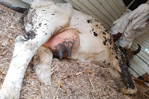
Clinical signs
Swollen edematous affected quarter hot to touch, pain on palpation of udder, fall in milk yield, pyrexia, anorexia, dullness, depression. Change in milk colour, flakes in milk, watery to reddish to thick creamy consistency of milk, milk with pus, milk with smell.
Acute mastitis
Sudden onset, high fever, anorexia, swollen udder hot to touch, enlarged supramammary lymph node, severe drop in milk yield. Curd to watery consistency of milk, edematous udder, sometimes death of animal observed due to severe toxaemia and per acute nature of infection.
Chronic mastitis
Animal with normal body temperature, appetite, fall in milk yield, shrinken udder, milk with pus or curdle or sticky consistency. Fibrosis and atrophy of affected quarter.
- Based on clinical signs like swelling of udder Hard swelling udder – acute mastitis Shrunken udder-chronic mastitis, change in consistency of milk like watery, reddish, yellowish and fall in milk yield.
- On physical examination of udder swollen quarter, hot on palpation in acute mastitis and asymmetrical udder.
- Ultrasonographic examination of udder to access the damage of affected quarter.
- History of change in udder consistency, change in milk composition from whitish to curdling, watery, with pus.
- Examination of milk in laboratory
- Normal pH of milk 6.4-6.8. So milk having ph above 7 indicates positive for mastitis.
- Bromothymol blue test.
- Mastrip indicator paper method.
- Bromocresol purple test
- Chloride test.
- Catalase test
- White side test.
- California mastitis test(CMT)
- NAGase test.
- Hotis test
- Electrical conductivity test
- Staining of milk by Newman,s stain.
- By checking bulk mill cell count and individual cow cell count.
- Culturing of milk in various bacteriological medium.
- Electrical Conductivity Meter Test
- Somatic cell count (SCC
- SCC through Digital
Treatment of mastitis
Treatment in mastitis is a challenging task. Here treatment is needed to stop the bacterial growth and to promote the milk yield. Isolation and identification of bacteria is more important along with antibiotic sensitivity test will gives the clear cut idea about therapeutic management. Some of intra-mammary preparations are given below.
- Intra mammary preparations
- Cefaperazone Sodium 250mg 10ml tube. Intra-mammary tube.
- Cefuroxime 250mg 3.5gm tube.
- Cefoquinome SO4 75mg 8gm tube.
- Azithromycin 300mg and Neomycin 250mg 5gm tube
- Ampicillin Sodium 75mg and Cloxacillin Sodium 200mg 10 ml tube.
- Colistin SO4 (5,00,000 IU) and Cloxacillin Sodium (200mg) 10ml tube
- Systemic antibiotic therapy as a synergistic effect for intra-mammary preparations.
- Anti-inflammatory preparations to reduce swelling, pain and pyrexia.
- To avoid the toxemia steroids should be given as per body weight.
- For the removel of debris and pus oxytocin should be given as per dose.
- To reduce the effect of histamine anti-histamine agents should be given.
- Fluid therapy should be given to reduce the toxemic effects and for early recovery.
- External applications to affected quarters to reduce swelling and pain.
- Vitamins and minerals supplementation.
Prevention and control
Mastitis is a managemental disease. It doesn’t have any vaccine. Earlier some of researchers tried on vaccination but results were not satisfactory. So prevention is better than cure applicable to mastitis. As the disease has more economical oriented proper hygiene and care should be taken in the farm. Clinical form of mastitis is about 5-10% where as subclinical mastitis is about 90-95%. In sub clinical mastitis udder will be normal, animal will be normal, milk appears normal, there will be low milk output, long duration of incidence and there will elevated somatic cell count more than 2,00,000 cells per ml of milk. It shows the importance of control of sub clinical mastitis. So early detection of sub clinical mastitis will be the first step for the control of clinical form of mastitis and to avoid the subsequent complications in animal health and economical losses in dairy industry. Some of effective control measures are given below.
- Clean and dry floor of the cattle shed.
- Keep animal clean.
- Vascularization of udder.
- Milker’s hygiene and complete milking practice.
- Time of milking should be same every day.
- Early isolation and treatment of affected cow.
- Regular cleaning and replacing of milking machine.
- Feeding of animal after milking.
- Allowing of calf to suckle after milking.
- Spraying or dipping of per chloride, weak iodophores, chlorhexidine solutions to teats after milking.
- Dry cow therapy.
- Isolation and prompt treatment of affected cows.
- Culling of chronically affected cows.
- Nutritional therapy to control the deficiency of vital nutrients.
- Periodical check up of cows for subclinical mastitis and prompt treatment.
- Avoid stress to animals.
- Strict quarantine for newly arrived animals.
- Regular changing of bedding material.
- Avoid wet floor in the farm.
- Animal with parturition complications like dystokia, uterine torsion, retention of placenta (ROP) should be isolated and treated promptly.

References
- Merck’s Veterinary Manual.
- Dr Prasanna Kumar notes on Bacterial diseases. Veterinary College Bidar Karnataka.
- Bovine mastitis: Theoretical and practical consideration in management. By P. Krishnamoorthy, M. Bhavana, Susweta Das Mitra, N. Krithiga, B.R. Shome.PD_ADMAS Hebbal Bangalore
| The content of the articles are accurate and true to the best of the author’s knowledge. It is not meant to substitute for diagnosis, prognosis, treatment, prescription, or formal and individualized advice from a veterinary medical professional. Animals exhibiting signs and symptoms of distress should be seen by a veterinarian immediately. |


Very nice 👌👌