Introduction
Coronaviridae family of viruses are the largest group of virus belonging to the order Nidovirales. Coronavirinae is one of the important subfamily of Coronaviridae further divided into four genera viz. alpha, beta, gamma and delta. These genera are sorted and divided based on phylogenetic clustering. Alphacoronavirus and Betacoronavirus can infect mammals. Gammacoronavirus and Deltacoronavirus mainly infect birds, fishes, and rarely cause disease in mammals (Woo et al., 2012; Cui et al., 2019).
Status in Indian Livestock
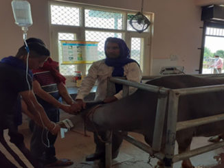
Animal coronavirus infections are widespread throughout the world including India. Bovine coronavirus is one of the commonest viral pathogens isolated from the cases of neonatal mortality in calves and winter dysentery in adult cattle. Rai and Singh, (1983) reported the isolation of coronavirus from neonatal calves suffering from pneumo-enteritis in India. Hansa et al. (2012) conducted screening of faecal samples recovered from diarrheic calves in Uttar Pradesh and reported 14-20% incidence rate for coronavirus. Kumar et al. (2013) performed detection of bovine coronavirus from diarrheic calves in states like Uttar Pradesh, Tamil Nadu and Karnataka. Shrivastava et al. (2018) performed testing of 38 faecal samples from calves died due to enteritis in Jabalpur region out of which 8 samples (21.05%) were detected positive for bovine coronavirus. Singh et al. (2019) conducted prevalence study of bovine coronavirus in dairy farms from Central (Madhya Pradesh and Chhattisgarh) and Northern (Uttar Pradesh) states of India. They observed a higher prevalence rate in North India (0.87%) as compared to Central India (0.83%). The prevalence of bovine coronavirus infection was higher in young calves below 2 months of age during the monsoon season.
Ganesan et al. (1990) detected the canine coronaviral enteritis for the first time in India. Twenty six (26) out of 900 samples (2.88%) were positive for coronavirus and about 58% of the affected dogs were belonging to the age group of below 6 months. Kaur et al. (2006) examined serum samples of dogs infected with haemorrhagic gastroenteritis. 4.45% of samples were detected positive for canine coronavirus (CCV). Deka et al. (2013) performed an epidemiological study on CCV infection in Assam and recorded a prevalence rate of 19.28%. Out of which 4.49% of the cases were suffering from mixed infection with canine parvovirus. Agnihotri et al. (2018) screened diarrheic faecal samples from dogs in Haryana. Out of 50 samples, 4 (8%) were positive for canine coronavirus whereas 50% of the samples were positive for canine parvovirus.
Barman et al. (2003) conducted a seroprevalence study on Transmissible gastroenteritis virus (TGV) in pigs from Assam. They detected prevalence rate of 39.4% for TGV infection. A high prevalence rate was reported in pigs reared under intensive system of farming.
Elankumaran et al. (1998) investigated tracheal swabs and serum samples procured from commercial layer flocks affected with infectious bronchitis virus (IBV) M41 and 793/B serotypes in Tamil Nadu. Kaul et al. (2009) collected post-mortem samples from birds affected with visceral gout in Jabalpur. They reported the isolation of IBV isolate with 95% sequence homology with Korean isolate and Chinese vaccine strains.
Patel et al. (2015) conducted isolation of five nephropathogenic IBV isolates from Gujarat. The study revealed 99-100% sequence homology among themselves and with Massachusetts vaccine strain. Gola et al. (2017) studied the prevalence of IBV in 10 commercial poultry farms in Uttarakhand. They observed mortality rate in the range of 5.4- 35.2%. Raja et al. (2019) reported the emergence of novel GI-24 IBV variant in India.
Genomic characterization
Coronaviruses are enveloped, pleomorphic (80-120nm size), non-segmented positive-sense RNA viruses having the largest genome size (33.5kb) among RNA viruses. Under the electron microscopy, club-shaped peplomers are seen arranged in a form of crown-like morphology so it is called as Corona. Mostly coronaviruses are having two types of peplomers such as spike protein and hemagglutinin-esterase glycoprotein. Among these two proteins, spike protein is longer and present in all of the coronaviruses. The spike protein is important for the tropism, pathogenic capability and virulence mechanism of virus (Lai and Holmes, 2001). Spike proteins have an ability to undergo mutation, which serves an important way to cause disease in a vast range of animals. Coronaviruses are able to undergo intraspecies recombination to evolve as virulent one but interspecies recombination rarely reported (Vijaykrishna et al., 2007; Gaunt et al., 2010). The viruses are subdivided into three major antigenic groups and known to infect multiple species of animals, birds and human beings. Group one is comprised of canine coronavirus, feline coronavirus, transmissible gastroenteritis virus and human. Bovine coronavirus, porcine hemagglutinating encephalomyelitis virus and murine hepatitis virus are members of group two. Group three includes infectious bronchitis virus and turkey coronavirus. ( Smith and Denison, 2012; Woo et al., 2012; Smith et al., 2013).
Canine coronavirus (CCoV) and feline coronavirus (FCoV)
Coronavirus can infect cat and dog resulting in feline infectious peritonitis infection (FIP) and canine enteric infection respectively. This feline and canine coronavirus belongs to the genus Alphacoronavirus. Some authors reported that mink and ferret coronavirus also comes under alphacoronavirus genus (Wise et al., 2010; Vlasova et al., 2011)
These viruses are further divided into two main genotypes such as type1 and type2 FCoV/CCoVs. Among these, type 2 FCoV is evolved from mixing of type1 FCoV and CCoVs (Herrewegh et al., 1998). More than 36% of infected dogs are having both types of genotypes and these viruses possess high capability of recombination and transmission to other species because of the recombination and mutation happening in the spike protein region. APN (Aminopeptidase) is a species-specific receptor for alphacoronavirus. Specifically feline APN serves as receptor for feline, porcine, canine and human coronavirus. Feline, porcine and human APN shows 78% similarity. But host-specific character is influenced by minor variations in APN (Benbacer et al., 1997; Tusell et al., 2007). This receptor sharing and small genetic changes are enough to cause zoonotic outbreaks with dangerous epidemic in the human population like SARS in 2003 (Peiris et al., 2003). Most cases of canine coronavirus are spread by oral contact with infected faeces and dog may also acquire the disease directly from infected dogs.
Worldwide enteric canine coronavirus infection is most common in dogs and coronoviral enteritis is also reported in wild dogs. This virus causes destruction of mature intestinal cells which leads to malabsorption and diarrhoea. But most commonly gastric disorder accompanied by soft, yellow-orange faeces is possible in subclinical infections (Erles et al., 2003). Pantropic strains (CCoV type II) of coronavirus have also been reported as a cause of severe systemic disease in dogs such as pyrexia, anorexia, depression, vomiting, diarrhoea, leukopenia, and neurologic signs of ataxia and seizures (Buonavoglia et al., 2006).
Feline infectious peritonitis (FIP) is caused by feline enteric coronavirus (FeCV), which is normally present in the digestive tract of cats. Any mutation or genetic changes leads to virulent strain (FIVP) formation and infect white blood cells, moreover spread all over the body of the cats (De Groot, 1995). FIP is a fatal, progressive and immune-mediated disease. It is prevalent <10% in coronavirus positive cats and causes disease in lesser than 2 years of old cat (kennady et al., 2002). Cat breeds like Abyssinian, Bengal, Himalayan and Rex are most susceptible to diseases. FIP is clinically classified in three distinctive forms. First one is wet form/effusive form, the second one is dry form/non-effusive or granulomatous form and mixed form is the third one. Among these forms, the effusive form is more common and death mostly occurs. In the second form of the disease, granulomatous lesions are noticed in the different organs including eyes and CNS. One form may transform into other forms. Along with these symptoms; fever, weight loss, anorexia, abdominal swelling and jaundice may also be reported (Riemer et al., 2016).
Bovine coronavirus (BCoV)
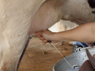
BCoV is categorized under the genus Betacoronavirus within the family Coronaviridae; also including HCoV-OC43, which causes respiratory contaminations in people, and the human pathogens SARS-CoV and MERS-CoV (Vijgen et al., 2006; Yang et al., 2015). This coronavirus is boundless in the livestock populace, bringing about monetary misfortunes to the dairy and beef industry throughout the world. The infection has been distinguished on all mainlands and there is serologic prevalence (>90%) that revealed most steers become presented to BCoV in the course of their life. It was the second most common respiratory infection after bovine herpesvirus disease (Kapil et al., 2008).
BCoV is transmitted by means of the faecal-oral or respiratory course. It can enter through respiratory and digestive system epithelial cells, nasal turbinates, trachea and lungs,. It is spread and shed by means of nasal secretions and faecal matters. (Clark, 1993; Park et al., 2007). Both in beef and dairy herds, BCoV is associated with calf diarrhoea, calf respiratory disease, winter dysentery in adult cattle and combined pneumonia and diarrhoea in calves and adults (Traven et al., 2001). Furthermore, this coronavirus can be clinically divided as bovine enteric or enteropathogenic coronaviruses (BECoV) and bovine respiratory coronaviruses (BRCoV) (Kapil and Goyal, 1995). Incidence of BcoV diarrhea may vary between different seasons. The viruses coming under the antigenic group 2 are able to jump between various species like from domestic to wildlife animals. The coronavirus infection status in small ruminants is less common (Park et al., 2007; Decaro et al., 2008).
Porcine coronavirus (PCoV)/Transmissible gastroenteritis virus (TGEV)
The coronaviruses infecting pigs are classified into Transmissible gastroenteritis virus, Haemagglutinating encephalomyelitis virus, porcine epidemic diarrhoea virus (PEDV), porcine respiratory coronavirus (PRCV) and Delta coronavirus (PDCoV). All the viruses except porcine respiratory coronavirus cause digestive disorders and TGEV is an economically important disease (Chen et al., 2014; Marthaler et al., 2014).
TGEV is a coronavirus which can infect pigs and cause significant economic losses in pig farming. This virus genus belongs to Alphacoronavirus and subgenus Tegacovirus. Aminopeptidase (APN) is a receptor for this virus (Woo et al., 2010; Fehr et al., 2015).
TGEV is a genetically closer relative of canine or feline coronavirus (Laude et al., 1990). The disease is most prevalent in colder temperatures. The infection spreads rapidly by aerosol route and by direct or indirect contact. It is a disease of small intestine that causes severe vomiting and profuse diarrhoea in pigs of all age groups. The incubation period of this virus is 18 hours.
Vomiting is the earliest sign of TGEV and followed by diarrhoea, which causes severe dehydration and saltwater. Electrolyte imbalance affects the cardiac function and finally animals end up with death. Piglets are most susceptible and death usually occur within 4 to 5 days. The herd mortality rate is less than 10-20%, but above 80% in piglets (Harris, 2019). Other enteric coronaviruses are emerging pathogens in developed countries causing epidemic diarrhoea in younger piglets. Among these, swine enteric coronaviruses are members of alpha and delta coronavirus, which are indistinguishable from TGEV virus; because they are serologically cross-reactive and the clinical features are not much significant (Ma et al., 2015; Zhang, 2016).
Equine coronavirus (ECoV)
Equine coronavirus (ECoV) is an emerging pathogen isolated in association with equine disease outbreak. It causes fever and enteric diseases in adult horses sporadically. In certain equine population, it causes substantial morbidity compared to expected mortality (Oue et al., 2012; Fieldling et al., 2015). Equine coronavirus is antigenically related to bovine coronavirus (Guy et al., 2000). Faeco-oral route of transmission is more common in equines. ECoV is unlikely to cause respiratory disease in horse but promptly it causes enteric diseases. It suggests that equine coronavirus is having a lesser affinity towards respiratory tract epithelium (Pusterla et al., 2015).
Infection of equine coronavirus is reported in horses older than two years of age but in foals, the disease is manifested as a combined infection with clostridium and rotavirus. The infection is acquired during winter months and the virus is shed from animals 3-4 days after clinical signs (Goodrich et al., 2018). The common clinical signs are anorexia, fever, profuse diarrhoea, colic-like symptoms and some neurological symptoms (ataxia, depression with encephalopathy). Death is rare but it can be possible because of secondary complications. Some of the animals recovered from the disease are also able to shed the virus (Giannitti et al. 2015).
Avian infectious bronchitis virus
Infectious bronchitis virus causes OIE-listed disease: avian infectious bronchitis (IB) in chickens. The disease renders poultry economic sector weak in the form of mortality, loss of body weight, increased feed conversion ratio and loss of production value (Kataria et al., 2005; Gowthaman et al., 2012). It is a member of gammacoronavirus and produces infection specific to respiratory, renal and reproductory system. IB is an acute, contagious disease and ubiquitously present all over the continents of broiler and layer type birds, which primarily starts with respiratory signs (Cavanagh & Gelb, 2008).
Airborne transmission is the most common route of spread but it can also transmit horizontally through faeces (Cavanagh and Naqi, 2003). The incubation period of this disease is 18-36 hours (Hofstad, 1984). The affected birds can carry the virus for more than months and shed large quantity of virus. About 30% mortality can occur due to secondary bacterial infections (M. gallisepticum, E. coli) and virulence status of serotypes are important for producing mortality in chickens (Rollier et al., 2000; Fabricant, 2000).
IBV affects all age groups of birds but severe infection is frequently seen in young ones. The common clinical manifestations are gasping, sneezing, respiratory rales, nasal discharge, ophthalmitis and sinusitis (Otuski et al., 1990). Broiler birds infected with nephrogenic form may help to cure respiratory form but renal symptoms like wet droppings, thirst and urolithiasis occur indefinitely (Cook et al., 2001; Arthur Sylvester, 2005).
Infection of day-old chicks leads to permanent damage to oviduct resulting in reduced egg production and quality (Mahesh Mahendran et al., 2005). Birds over 8 to 16 weeks of age exhibit only mild respiratory symptoms. IB viruses produce some lung lesions with caseous exudates in congested lungs. In the renal system, it produces swollen ureter with urate accumulation seen in kidneys. Reproductive lesions like underdeveloped blind sac projecting from cloaca, atrophy of isthmus-magnum junction, cystic dilatation of oviduct are noted (Cavanaghand Naqi, 2003; Bhattacharjee and Jones, 1997). More specifically this virus produces pectoral oedema and haemorrhages (DhinakarRaj and Jones, 1996).
Zoonotic values
The two important pathogenic human coronaviruses (HCoV’s), SARS-CoV (severe acute respiratory syndrome), MERS-CoV (Middle East respiratory syndrome) and SARS2-CoV (severe acute respiratory syndrome 2) have been identified as zoonotic infections by various research workers. Bats play a pivotal role in the transmission of HCoVs in the form of natural reservoirs including the recent pathogen 2019-nCoV responsible for the global pandemic. Bats harbour the viral agents inside their body without development of any pathogenic conditions in themselves. Among the wild animals, Civet cats, racoons, pangolin and dogs are reported as the carriers for SARS-CoV infection to human beings (Guan et al., 2003; WHO, 2020).
Various studies were conducted over the period of time reported dromedary camels from the Middle-East and African regions serve as primary infection source of MERS-CoV to the human beings (Ahmed et al., 2018). Saquib et al. (2017) suggested the role of dromedary camels in the pathogenesis of human SARS-Cov infection in Punjab province of Pakistan. A study conducted by Sikkema et al. (2017) in Qatar, Saudi Arabia emphasized that exposure to dromedary camels serves as a prime risk factor for the MERS-CoV infection among the camel workers.
Diagnosis of Animal coronaviruses
Diagnosis of coronavirus can be done with many techniques from the isolation of virus to serology, immunopathology and next-generation sequencing technology. Nasal swab and diarrhoea samples are used for virus isolation and antigen detection. PCR and Reverse transcriptase PCR are the most sensitive methods for the detection of viral RNA (Oma et al., 2016; Roh et al., 2014). In serological tests, ELISA, HI test, agar gel precipitation test and viral neutralisation assays are used to identify corona-specific antibodies in serum of the animals. Analysis of peritoneal effusion fluid is the most sensitive method for detecting antigen in felines (Felton and Hartman, 2019). For IBV disease lung, oviduct, kidney can be collected and used for antigen detection by immunohistochemistry. Genetic diversity of virus can be identified with sequencing spike protein gene and phylogenetic analysis (Valastro et al., 2016).
Micro neutralization assay is confirmatory test to detect neutralizing antibodies in animals and humans. Real-time reverse-transcription polymerase chain reaction (rRT-PCR) test is most sensitive (>80%) for diagnose viral RNA and results will get within two hours. High throughput style COVID-19 diagnostic instruments, such as Abbott’s m2000 Real Time system is FDA approved High throughput covid19 diagnostics, which use molecular PCR detection technology. It can diagnose 470 test per day. Rapid diagnostic methods such as Antigen and Antibody detection based on paper strip is mostly used in pandemic times for point of care diagnosis.
Prevention and control of Animal coronaviruses
Infected animals should be isolated separately and maintained in quarantine shed with stress-free conditions. Animal excretions contain many viruses which spread disease in other animals. The virus can spread directly and indirectly through contaminated inanimate objects. Disinfection (0.5% chlorine solution, peracetic or peroxyacetic acid at concentrations of 500−2000 mg/L, 70% ethyl alcohol, sodium hypochlorite at 0.5%) of the animal shed and equipments are mandatory to kill the virus. Landfills are used to remove the animals’ excretions and bedding materials. Composting of manures may decontaminate infectious pathogen. Bovine coronaviruses are controlled by vaccinating dam with live vaccine ATCvet code QI02 (WHO Collaborating Centre for Drug Statistics Methodology, Intra nasal route). It will help in the transfer of immunity to calves by colostrum. For controlling IBV many live vaccines and inactivated vaccines are used in the poultry industry (Britton and Cavanagh, 2007). Prevention of secondary bacterial infection is an imperative step to avoid high rate of mortality. Application of formaldehyde, quaternary ammonium compounds and chlorine releasing agents are commonly used to eliminate the virus. But concentration and contact time of disinfectant is an important factor to kill the virus (Sjaak de Wit et al., 2011). Rapid diagnostic measures/kits, effective vaccines and novel therapeutics are essential to control coronavirus infection among animals. Beside these, suitable biosecurity measurements and proper management are necessary to overcome coronavirus infection in animals.
Reference
- Agnihotri, D., Singh, Y., Batra, K., Jain, V.K., Kumar, A., Kumar, T. and Maan, S., 2018.
- Ahmed, A.E., Al-Jahdali, H., Alshukairi, A.N., Alaqeel, M., Siddiq, S.S., Alsaab, H., Sakr, E.A., Alyahya, H.A., Alandonisi, M.M., Subedar, A.T. and Aloudah, N.M., 2018. Early identification of pneumonia patients at increased risk of Middle East respiratory syndrome coronavirus infection in Saudi Arabia. International Journal of Infectious Diseases, 70, pp.51-56.
- Barman, N.N., Barman, B., Sarma, D.K. and Pensaert, M.B., 2003. Prevalence of rotavirus, transmissible gastroenteritis virus and porcine epidemic diarrhoea virus antibodies in pigs of Assam, India. Indian Journal of Animal Sciences (India).
- Benbacer, L., Kut, E., Besnardeau, L., Laude, H. and Delmas, B., 1997. Interspecies aminopeptidase-N chimeras reveal species-specific receptor recognition by canine coronavirus, feline infectious peritonitis virus, and transmissible gastroenteritis virus. Journal of virology, 71(1), pp.734-737.
- Bhattacharjee, P.S. and Jones, R.C., 1997. Susceptibility of organ cultures from chicken tissues for strains of infectious bronchitis virus isolated from the intestine. Avian Pathology, 26(3), pp.553-563.
- Buonavoglia, C., Decaro, N., Martella, V., Elia, G., Campolo, M., Desario, C., Castagnaro, M. and Tempesta, M., 2006. Canine coronavirus highly pathogenic for dogs. Emerging infectious diseases, 12(3), p.492.
- Cavanagh, D. and Naqi, S.A., 2003. Infectious bronchitis. Diseases of poultry, 11, pp.101-119.
- Chen, X., Yang, X., Zheng, Y., Yang, Y., Xing, Y. and Chen, Z., 2014. SARS coronavirus papain-like protease inhibits the type I interferon signaling pathway through interaction with the STING-TRAF3-TBK1 complex. Protein & cell, 5(5), pp.369-381.
- Clark, M.A., 1993. Bovine coronavirus. British Veterinary Journal, 149(1), pp.51-70.
- Cook, J.K.A., Chesher, J., Baxendale, W., Greenwood, N., Huggins, M.B. and Orbell, S.J., 2001. Protection of chickens against renal damage caused by a nephropathogenic infectious bronchitis virus. Avian Pathology, 30(4), pp.423-426.
- Cui, J., Li, F. and Shi, Z.L., 2019. Origin and evolution of pathogenic coronaviruses. Nature reviews Microbiology, 17(3), pp.181-192.
- De Wit, J.J., Cook, J.K. and Van der Heijden, H.M., 2011. Infectious bronchitis virus variants: a review of the history, current situation and control measures. Avian Pathology, 40(3), pp.223-235.
- Deka, D., Phukan, A. and Sarma, D.K., 2013. Epidemiology of parvovirus and coronavirus infections in dogs in Assam. Indian Vet. J, 90(9), pp.49-51.
- Elankumaran, S., Balachandran, C., Chandran, N.D.J., Roy, P., Albert, A. and Manickam, R., 1999. Serological evidence for a 793/B related avian infectious bronchitis virus in India. Veterinary record, 144(11), pp.299-300.
- Erles, K., Toomey, C., Brooks, H.W. and Brownlie, J., 2003. Detection of a group 2 coronavirus in dogs with canine infectious respiratory disease. Virology, 310(2), pp.216-223.
- Fehr, A.R. and Perlman, S., 2015. Coronaviruses: an overview of their replication and pathogenesis. In Coronaviruses(pp. 1-23). Humana Press, New York, NY.
- Fielding, C.L., Higgins, J.K., Higgins, J.C., McIntosh, S., Scott, E., Giannitti, F., Mete, A. and Pusterla, N., 2015. Disease associated with equine coronavirus infection and high case fatality rate. Journal of veterinary internal medicine, 29(1), pp.307-310.
- Ganesan, P.I., Ramadass, P. and Gunaseelan, L., 1990. Detection of canine coronavirus enteritis. Indian Veterinary Journal, 67(11).
- Gaunt, E.R., Hardie, A., Claas, E.C., Simmonds, P. and Templeton, K.E., 2010. Epidemiology and clinical presentations of the four human coronaviruses 229E, HKU1, NL63, and OC43 detected over 3 years using a novel multiplex real-time PCR method. Journal of clinical microbiology, 48(8), pp.2940-2947.
- Giannitti, F., Diab, S., Mete, A., Stanton, J.B., Fielding, L., Crossley, B., Sverlow, K., Fish, S., Mapes, S., Scott, L. and Pusterla, N., 2015. Necrotizing enteritis and hyperammonemic encephalopathy associated with equine coronavirus infection in equids. Veterinary pathology, 52(6), pp.1148-1156.
- Gola, S., Shukla, S.K., Shekhar, S. and Kumar, M., 2017. Prevalence Serodiagnosis and Histopathological Changes in Field Cases of Infectious Bronchitis in Chickens. Int. J. Curr. Microbiol. App. Sci, 6(8), pp.1591-1597.
- Goodrich, E.L., Mittel, L.D., Glaser, A., Ness, S.L., Radcliffe, R.M. and Divers, T.J., 2018. Novel findings from a beta coronavirus outbreak on an American Miniature Horse breeding farm in upstate New York. Equine Veterinary Education.
- Gowthaman, V., Singh, S.D., Dhama, K., Barathidasan, R., Kumar, M.A., Desingu, P.A., Mahajan, N.K. and Ramakrishnan, M.A., 2012. Fowl adenovirus (FAdV) in India: Evidence for emerging role as primary respiratory pathogen in chickens. J. Biol. Sci, 15(18), pp.900-903.
- Guan, Y., Zheng, B.J., He, Y.Q., Liu, X.L., Zhuang, Z.X., Cheung, C.L., Luo, S.W., Li, P.H., Zhang, L.J., Guan, Y.J. and Butt, K.M., 2003. Isolation and characterization of viruses related to the SARS coronavirus from animals in southern China. Science, 302(5643), pp.276-278.
- Hansa, A., Rai, R.B., Wani, M.Y. and Dhama, K., 2012. Patholology and diagnosis of corona virus infection in bovine. Indian Journal of Veterinary Pathology, 36(2), pp.129-135.
- Herrewegh, A.A., De Groot, R.J., Cepica, A., Egberink, H.F., Horzinek, M.C. and Rottier, P.J., 1995. Detection of feline coronavirus RNA in feces, tissues, and body fluids of naturally infected cats by reverse transcriptase PCR. Journal of clinical microbiology, 33(3), pp.684-689.
- Herrewegh, A.A., Smeenk, I., Horzinek, M.C., Rottier, P.J. and de Groot, R.J., 1998. Feline coronavirus type II strains 79-1683 and 79-1146 originate from a double recombination between feline coronavirus type I and canine coronavirus. Journal of virology, 72(5), pp.4508-4514.
- Hofstad, M. S. (1984). Avian infectious bronchitis. In “Diseases of Poultry” (B. W. Calnek, H. J. Barnes, C. W. Beard, W. M. Reid, and H. W. Yoder, Eds.), 8th ed., pp. 429–443. Iowa State Univ. Press, Ames, IA
- Kumar, S.S., Rai, R.B., Dhama, K., Ranganath, G.J., Saminathan, M., Wani, M.Y. and Saravanan, R., 2013. Detection of bovine coronavirus in calf diarrheic samples by indirect antigen capture ELISA and RT-PCR. Research Opinions in Animal and Veterinary Sciences, 3(8), pp.225-234.
- Kumar, V., Jung, Y.S. and Liang, P.H., 2013. Anti-SARS coronavirus agents: a patent review (2008–present). Expert opinion on therapeutic patents, 23(10), pp.1337-1348.
- Lai and Holmes, 2001 M.M.C. Lai, K.V. Holmes Coronaviridae: The Viruses and Their Replication in Fundamental Virology (4th Edition), Lippincott Raven, Philadelphia (2001)
- Tråvén, K. Näslund, L. Linde, B. Linde, A. Silván, C. Fossum, K.O. Hedlund, B. Larsson Experimental reproduction of winter dysentery in lactating cows using BCV – Comparison with BCV infection in mild-fed calves Veterinary Microbiology, 81 (2001), pp. 127-151
- Ma, Y., Wu, L., Shaw, N., Gao, Y., Wang, J., Sun, Y., Lou, Z., Yan, L., Zhang, R. and Rao, Z., 2015. Structural basis and functional analysis of the SARS coronavirus nsp14–nsp10 complex. Proceedings of the National Academy of Sciences, 112(30), pp.9436-9441.
- Marthaler, D., Jiang, Y., Collins, J. and Rossow, K., 2014. Complete genome sequence of strain SDCV/USA/Illinois121/2014, a porcine deltacoronavirus from the United States. Genome Announc., 2(2), pp.e00218-14.Molecular Detection Based Epidemiology of Canine Parvovirus and Canine Coronavirus Infection in Diarrheic Dogs in Haryana. Journal of Animal Research, 8(3), pp.367-374.
- Oma, V.S., Tråvén, M., Alenius, S., Myrmel, M. and Stokstad, M., 2016. Bovine coronavirus in naturally and experimentally exposed calves; viral shedding and the potential for transmission. Virology journal, 13(1), p.100.
- Otsuki, K., Huggins, M.B. and Cook, J.K., 1990. Comparison of the susceptibility to avian infectious bronchitis virus infection of two inbred lines of white leghorn chickens. Avian Pathology, 19(3), pp.467-475.
- Park, S.J., Kim, G.Y., Choy, H.E., Hong, Y.J., Saif, L.J., Jeong, J.H., Park, S.I., Kim, H.H., Kim, S.K., Shin, S.S. and Kang, M.I., 2007. Dual enteric and respiratory tropisms of winter dysentery bovine coronavirus in calves. Archives of virology, 152(10), pp.1885-1900.
- Patel, B.H., Bhimani, M.P., Bhanderi, B.B. and Jhala, M.K., 2015. Isolation and molecular characterization of nephropathic infectious bronchitis virus isolates of Gujarat state, India. Virusdisease, 26(1-2), pp.42-47.
- Pusterla, N., Vin, R., Leutenegger, C., Mittel, L.D. and Divers, T.J., 2016. Equine coronavirus: An emerging enteric virus of adult horses. Equine Veterinary Education, 28(4), pp.216-223.
- Rai, R.B. and Singh, N.P., 1983. Isolation of coronavirus from neonatal calves with pneumoenteritis in India. Hansa, A., Rai, R.B., Dhama, K. and Wani, M.Y., 2012. ELISA and RT-PCR based detection of bovine coronavirus in Northern India. Asian Journal of Animal and Veterinary Advances, 7(11), pp.1120-1129.
- Raj, G.D. and Jones, R.C., 1996. An in vitro comparison of the virulence of seven strains of infectious bronchitis virus using tracheal and oviduct organ cultures. Avian Pathology, 25(4), pp.649-662.
- Raja, A., Dhinakar Raj, G. and Kumanan, K., Emergence of variant avian infectious bronchitis virus in India.
- Riemer, F., Kuehner, K.A., Ritz, S., Sauter-Louis, C. and Hartmann, K., 2016. Clinical and laboratory features of cats with feline infectious peritonitis–a retrospective study of 231 confirmed cases (2000–2010). Journal of feline medicine and surgery, 18(4), pp.348-356.
- Roh, H.J., Jordan, B.J., Hilt, D.A., Ard, M.B. and Jackwood, M.W., 2015. Hatchery spray cabinet administration does not damage avian coronavirus infectious bronchitis virus vaccine based on analysis by electron microscopy and virus titration. Avian diseases, 59(1), pp.149-152.
- Rollier, C., Charollois, C., Jamard, C., Trepo, C. and Cova, L., 2000. Early life humoral response of ducks to DNA immunization against hepadnavirus large envelope protein. Vaccine, 18(27), pp.3091-3096.
- Saqib, M., Sieberg, A., Hussain, M.H., Mansoor, M.K., Zohaib, A., Lattwein, E., Müller, M.A., Drosten, C. and Corman, V.M., 2017. Serologic evidence for MERS-CoV infection in dromedary camels, Punjab, Pakistan, 2012–2015. Emerging infectious diseases, 23(3), p.550.
- Shrivastava, D.P., Swamy, M. and Dubey, A., 2018. Enteric Pathology of cattle calves naturally infected with corona virus in Jabalpur region. Indian Journal of Veterinary Pathology, 42(1), pp.64-67.
- Singh, S., Singh, R., Singh, K.P., Singh, V., Malik, Y.P.S., Kamdi, B., Singh, R. and Kashyap, G., 2019. Prevalence of bovine coronavirus infection in organized dairy farms of Central and North regions, India. Biological Rhythm Research, pp.1-7.
- Smith, E.C. and Denison, M.R., 2012. Implications of altered replication fidelity on the evolution and pathogenesis of coronaviruses. Current opinion in virology, 2(5), pp.519-524.
- Smith, E.C., Blanc, H., Vignuzzi, M. and Denison, M.R., 2013. Coronaviruses lacking exoribonuclease activity are susceptible to lethal mutagenesis: evidence for proofreading and potential therapeutics. PLoS pathogens, 9(8).
- Sylvester, S.A., Dhama, K., Kataria, J.M., Rahul, S. and Mahendran, M., 2005. Avian infectious bronchitis: A review. Indian Journal of Comparative Microbiology, Immunology and Infectious Diseases, 26(1), pp.1-14.
- Tusell, S.M., Schittone, S.A. and Holmes, K.V., 2007. Mutational analysis of aminopeptidase N, a receptor for several group 1 coronaviruses, identifies key determinants of viral host range. Journal of virology, 81(3), pp.1261-1273.
- Valastro, V., Holmes, E.C., Britton, P., Fusaro, A., Jackwood, M.W., Cattoli, G. and Monne, I., 2016. S1 gene-based phylogeny of infectious bronchitis virus: an attempt to harmonize virus classification. Infection, Genetics and Evolution, 39, pp.349-364.
- Vijaykrishna D., Smith G. J., Zhang J. X., Peiris J. S., Chen H., Guan Y. (2007). Evolutionary insights into the ecology of coronaviruses. J Virol 81, 4012–4020 10.1128/JVI.02605-06
- Vijgen, L., Keyaerts, E., Lemey, P., Maes, P., Van Reeth, K., Nauwynck, H., Pensaert, M. and Van Ranst, M., 2006. Evolutionary history of the closely related group 2 coronaviruses: porcine hemagglutinating encephalomyelitis virus, bovine coronavirus, and human coronavirus OC43. Journal of virology, 80(14), pp.7270-7274.
- Vlasova, A.N., Halpin, R., Wang, S., Ghedin, E., Spiro, D.J. and Saif, L.J., 2011. Molecular characterization of a new species in the genus Alphacoronavirus associated with mink epizootic catarrhal gastroenteritis. The Journal of general virology, 92(Pt 6), p.1369.
- Wise, A.G., Kiupel, M., Garner, M.M., Clark, A.K. and Maes, R.K., 2010. Comparative sequence analysis of the distal one-third of the genomes of a systemic and an enteric ferret coronavirus. Virus research, 149(1), pp.42-50.
- Woo, P.C., Huang, Y., Lau, S.K. and Yuen, K.Y., 2010. Coronavirus genomics and bioinformatics analysis. viruses, 2(8), pp.1804-1820.
- Woo, P.C., Lau, S.K., Li, K.S., Tsang, A.K. and Yuen, K.Y., 2012. Genetic relatedness of the novel human group C betacoronavirus to Tylonycteris bat coronavirus HKU4 and Pipistrellus bat coronavirus HKU5. Emerging microbes & infections, 1(1), pp.1-5.
- Oue, R. Ishihara, H. Edamatsu, Y. Morita, M. Yoshida, M. Yoshima, S. Hatama, K. Murakami, T. Kanno Isolation of an equine coronavirus from adult horses with pyrogenic and enteric disease and its antigenic and genomic characterization in comparison with the NC99 strain Veterinary Microbiology, 150 (2011), pp. 41-48
- Yang, D. and Leibowitz, J.L., 2015. The structure and functions of coronavirus genomic 3′ and 5′ ends. Virus research, 206, pp.120-133.
- Zhang, Q. and Yoo, D., 2016. Immune evasion of porcine enteric coronaviruses and viral modulation of antiviral innate signaling. Virus research, 226, pp.128-141.
|
The content of the articles are accurate and true to the best of the author’s knowledge. It is not meant to substitute for diagnosis, prognosis, treatment, prescription, or formal and individualized advice from a veterinary medical professional. Animals exhibiting signs and symptoms of distress should be seen by a veterinarian immediately. |


An extensive review.
Excellent depiction of corona