Introduction
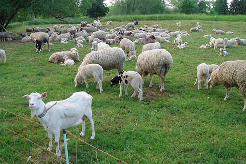
Peste des Petits Ruminants (PPR) is a devastating viral disease of small ruminants for decades that predominantly target sheep and goats. The disease also has capacity to infect several other animal species like cattle, buffalo, pig, and camel and wild ruminants. It is infectious as well as contagious disease thus, can spread very rapidly among the flock of animals. The disease is very serious causes major socio-economic imbalance and has no transboundary restrictions. In a country like India bearing the world’s largest livestock population where a large number of people are dependent on animal husbandry for their income, diseases like PPR may have damaging effects on livelihood as well as the economy. PPR is also famous with some other names such as goat plague, a plague of small ruminants, contagious pustular stomatitis, and ovine rinderpest.

The cause
The causative agent of PPR is the Peste des Petits Ruminants Virus from the Morbillivirus genus of the Paramyxoviridae family. It is a small negative sense, single-stranded RNA virus with a genome size of 15.9 kb. The virus is having a pleomorphic shape with size ranging from 150 to 700 nm. PPR virus is very fragile, susceptible to heat, ultraviolet light, and dehydration. It is unable to survive for long period outside the host. It has a half-life of approximately 22 minutes at 56 °C and 3.3 hours at 37° C. It is stable between pH ranges of 5.8 to 9.5 at normal temperature but gets degraded at highly acidic (below 4) and highly alkaline (above 11) pH. . In fresh meat, the virus takes relatively longer time for inactivation. Also, it can survive up to 72 hours in shaded conditions.
Epizootiology and transmission
PPR disease has become endemic in most of countries of Africa, the Middle East, and Asia including China and India. In comparison to sheep, goats are found to be more susceptible and show an extreme form of clinical disease. Besides, goats have a low recovery rate than sheep.
Fortunately, the virus possesses only one serotype, but based on different strains it is differentiated into four distinct lineages. The base for the lineage classification is the fusion (F) gene or the nucleoprotein (N) gene sequence. Lineage I is common in Central Africa; lineage II in West Africa; whereas lineage III occurs in East Africa, the Middle East, and southern India and lineage IV is dominant in different parts of Asia. All the isolates from India are members of lineage IV except the ‘Arasur virus’ reported from the Tamil Nadu region which belongs to lineage III.
Domestic animals infected with the virus are the foremost source of the PPR virus. Direct contact between susceptible and infected animals causes airborne transmission which is the main mode of transmission. During an early stage of infection, all bodily secretions and excretions are flooded with virus load. While coughing and sneezing, the virus is ejected in the environment as droplets and contaminate the surrounding which favours virus transmission. As it is a contagious virus, animals housed together with infected one remains at higher risk. Movement of infected animals and trade helps in spreading the disease in non-infected areas. PPR virus does not spread through vertical transmission, i.e, from infected dam to its foetus during pregnancy
Economic Impact of PPR
FAO and OIE consider PPR as one of the most damaging transboundary diseases which causes major health as well as economic losses to small ruminant production. As per 20th Livestock census, 2019, India, has a huge animal population containing 148.88 million goats and 74.26 million sheep, considering this, PPR is a potential havoc for small ruminants with a mortality rate of around 50% or more. The presence of disease limits animal trade and livestock products which ultimately affects the returns supposed to be obtained from animals. Being most susceptible, kid mortality can reach up to100% in severe cases. Estimated annual direct and indirect financial losses due to PPRV are up to 1,800 million Indian Rupees (US$ 39 million). Direct annual losses are between USD 1.2 and 1.7 billion. Only the cost for PPR vaccination itself ranges between USD 270 and 380 million.
Pathogenesis and clinical signs
PPR virus gains entry inside the host by attachment with SLAM and nectin 4 receptors. After entry, virus initially replicates within the nasopharyngeal/respiratory epithelium. In the first 2-4 days after infection virus lodge within the lymph nodes. Subsequently, it gets disseminated causing viremia. Morbilliviruses have strong tropism or affinity towards lymphoid tissues. PPRV is also shows epitheliotropic nature like other morbilliviruses. It causes the destruction of lymphoid tissues leading to leucopenia and immunosuppression. Clinical signs of disease are visible after an incubation period of 4-6 days. In the acute case, the sudden onset of fever with 40°C to 41°C body temperature can be witnessed., animals become depressed, sleepy and hair are rough and erected. During the initial phase, a clear watery discharge appears from the eyes, nose, and mouth which consequently becomes thick and yellow. The discharges cause matting of the eyelids, obstruction of the nose, and difficult breathing. Sever form of pneumoenteritis develops producing dehydration and pasting of hindquarters with diarrhoea. Erosion of epithelium on the outer and inner side of the lips, lower gums produce ulcerative necrotic lesions in the oral cavity and the gastrointestinal tract. Due to acute pulmonary congestion, animals may succumb to death within a week. In some cases, chronic infection occurs with pneumonia which may get complicated further. Ultimately, the fate of the infection is determined by the immune response developed by an individual.
Diagnosis of PPR
Based on clinical signs, species affected and post-mortem lesions diagnosis can be made. But it should be differentiated from other similar symptoms producing diseases such as rinderpest, foot and mouth disease, contagious ecthyma, bluetongue, etc.
Post-mortem lesions
Onsite primary diagnosis can be made on the basis of PM lesions where laboratory facility is not easily available. Animals suspected for PPR may show characteristic inflammatory and necrotic lesions in the oral cavity and gastrointestinal tract upon PM examination. Few times lesions can be observed on the vulval and vaginal mucous membranes as well. Carcass appears emaciated or dehydrated along with crusted scabs on lips. Erosive lesions in the abomasum along with haemorrhages in duodenum and ileum can be found. Zebra stripes” or “tiger stripes” are the most prominent lesions in the large intestine and are considered as confirmatory or pathognomic lesions of PPR. Bronchopneumonia, congestion of lung, and petechial haemorrhages in the respiratory tract may present. Lymph nodes become swollen, with oedematous and necrotic changes can be observed. The liver and spleen may have necrotic lesions.
Laboratory diagnosis
Laboratory testing provides a confirmatory diagnosis. Virus neutralization test (VNT) is the OIE recommended test as a prerequisite for international trade of animals. Virus isolation is considered a gold standard for disease diagnosis. Molecular assays including RT-PCR and real-time RT-PCR targeting F and N gene of PPR are more sensitive methods for diagnosis. Antibody detection through serological tests like agar gel immuno-diffusion test (AGID)/AGPT, counter immuno-electrophoresis (CIE) and different types of ELISA are also available. Competitive enzyme-linked immunosorbent assay (c-ELISA) is widely used in serological diagnosis of PPRV infection as it is highly sensitive and specific. Rapid diagnosis can be done with Immuno-capture ELISA using monoclonal antibodies.
Prevention of PPR- Vaccination
Vaccination is the most compelling way to prevent and control PPR. Indian originated live attenuated vaccine, sungri/96 developed by Indian Veterinary Research Institute, Mukteswar is a commercialized vaccine used for mass vaccination. The only drawback of this commercial sungri/96 vaccine is thermo-sensitivity. Its life is around 1 year at 4°C. Hence an effective cold chain is needed to maintain its efficacy under the field condition which is not an easy task. This demands the need for the thermostable vaccine for the robust use in field conditions. The other two vaccines developed by TANUVAS are Arasure/87 from sheep origin and Coimbatore/97 from goat origin are also available for vaccination. Animals should be vaccinated after 3–4 months of birth when maternal antibodies get vanished. Combined vaccines consisting of PPR/Sungri/96 and SPPV- Romanian Fanar as well as PPRV-Sungri/96 and GTPV-Uttarkashi strains are also available to control both capripox and PPR. Recombinant vaccine using F or H genes of PPRV and capripoxvirus as vector (rCPV- PPR) shows protection in the case of both PPR and capripox disease. At present, the vaccine to differentiate infected from vaccinated animals (DIVA) is not available for PPR, which is the need of time for an effective eradication strategy.
Control and eradication strategy
Though vaccination is the major step toward disease control, other necessary precautions are must to prevent disease spread. Restriction of affected animal movement to avoid secondary cases production and segregation of sick animals from healthy ones are the foremost important actions to be taken in the affected flock. Carcasses of infected animals should dispose of by incineration or deep burial to avoid possible transmission. Proper disposal of contact material and decontamination of the surrounding is an important step to control further spread. On the strategic front, FAO and OIE have launched the global campaign for PPR eradication by 2030. Global Strategy for the eradication of PPR is a 15-year program of three five-year phases with estimated global costs of $ 996 Million for 5 years. In India, PPR-Control Programme was initiated during 11th five-year plan of and has now been enforced throughout the country by Government of India for mass vaccination of eligible sheep & goat population at free of cost in all the States & Union Territories.
Summary and Conclusion
Peste Des Petits Ruminants (PPR) is an OIE notifiable and transboundary disease-causing a constant threat to the countries all over the world raising small ruminant population. The disease is more severe in goats than sheep. Among the four lineages, lineage IV is circulating in Asia. The disease causes severe immunosuppression and produce respiratory and gastrointestinal tract symptoms with significant mortality rate and resultant financial losses. Various diagnostic tests are available to detect antigen as well as antibodies for PPRV. The disease is highly contagious so separation of infected animals and curtailment of animal trade is the best way to prevent further transmission. A live attenuated vaccine is available which is effective to prevent the disease. Vaccination of all animals at the age of 3-4 months provides immunity for 3 years with one shot of vaccine. Efforts for the global eradication of the disease with all the applied strategies will help to curb the disease from the world to safeguard the lives of animals and prevent the possible financial losses.
References
- 11th five-year plan of 2002-2007, Government of India, New Delhi.
- 20th livestock census of India. 2029. Department of animal husbandry, dairy and fisheries, Government of India, New Delhi.
- Bailey, D., Banyard, A., Dash, P., Ozkul, A., & Barrett, T. (2005). Full genome sequence of peste des petits ruminants virus, a member of the Morbillivirus genus. Virus research, 110(1-2), 119-124.
- Balamurugan, V., Sen, A., Venkatesan, G., Rajak, K. K., Bhanuprakash, V., & Singh, R. K. (2012). Study on passive immunity: Time of vaccination in kids born to goats vaccinated against Peste des petits ruminants. Virologica Sinica, 27(4), 228-233.
- Bardhan, D., Kumar, S., Anandsekaran, G., Chaudhury, J. K., Meraj, M., Singh, R. K., … & Mishra, V. (2017). The economic impact of peste des petits ruminants in India. Revue scientifique et technique (International Office of Epizootics), 36(1), 245.
- Baron, M. D., Diallo, A., Lancelot, R., & Libeau, G. (2016). Peste des petits ruminants virus. In Advances in virus research (Vol. 95, pp. 1-42). Academic Press.
- Diallo, A., Minet, C., Le Goff, C., Berhe, G., Albina, E., Libeau, G., & Barrett, T. (2007). The threat of peste des petits ruminants: progress in vaccine development for disease control. Vaccine, 25(30), 5591-5597.
- Gibbs, P. J., Taylor, W. P., Lawman, M. J., & Bryant, J. (1979). Classification of peste des petits ruminants virus as the fourth member of the genus Morbillivirus. Intervirology, 11(5), 268-274.
- Khan, H. A., Siddique, M., Abubakar, M., & Ashraf, M. (2008). The detection of antibody against peste des petits ruminants virus in sheep, goats, cattle and buffaloes. Tropical animal health and production, 40(7), 521-527.
- Singh, R. K., Balamurugan, V., Bhanuprakash, V., Sen, A., Saravanan, P., & Yadav, M. P. (2009). Possible control and eradication of peste des petits ruminants from India: technical aspects. Vet Ital, 45(3), 449-462.
- Taylor, W. P. (1979). Protection of goats against peste-des-petits-ruminants with attenuated rinderpest virus. Research in veterinary science, 27(3), 321-324.


Very nicely explained with scientific knowledge..