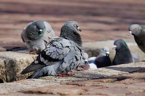
Pigeons were evolving to be one of the most common pets in the world. they were present in every part of the world except the colder regions. It comes under the Columbidae family which includes both doves and pigeons, the smaller ones are usually called as the doves. In many states like Tamil nadu and Kerala pigeons are even used for games, so its very essential to know the diseases of pigeons.
In this article I tried to explain about the common parasites affecting the digestive system of the pigeons. For easy understanding and remembrance I have divided the parasites based on the part of the digestive system which is affected.
PARASITES AFFECTING DIGESTIVE SYSTEM OF PIGEONS:
PARASITES AFFECTING CROP AND PROVENTRICULUS:
1. Trichomonas gallinae
Trichomonas gallinae is extremely common in domestic pigeons and often causes serous lesions.
Common name: canker, frounce, roup.
Predilection site: pharynx, esophagus, crop and proventriculus.
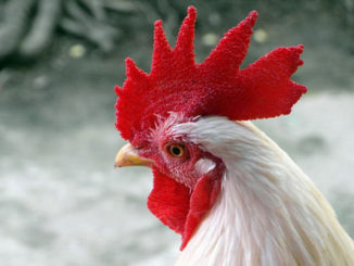
Hosts: : Pigeon, turkey, chicken, raptors.
Description: The body is elongate, ellipsoidal or pyriform, t. The undulating membrane does not reach the posterior end of the body and a free posterior flagellum is absent, There is a crescent‐shaped pelta anterior to the axostyle and there is no chromatic ring at its point of emergence. The parabasal body is hook‐shaped and has a parabasal filament and the costa is a very fine rod running three‐quarters the length of the body
Pathogenesis: The domestic pigeons is the primary host, but the parasite has been found in the birds of prey that feed on pigeons. Adult pigeons which are previously infected may act as symptomless carriers. Injection of plasma from infected birds also confers immunity. Trichomonas is essentially a disease of young birds. 80-90% of the adults are infected but shows no signs of the disease.
Clinical signs: Severely affected birds lose weight, stand huddled with ruffled feathers and may fall over when forced to move. Yellow necrotic lesions are present in the mouth, oesophagus and crop of pigeon squabs and a greenish fluid containing large numbers of trichomonads may be found in the mouth. The condition is often fatal.
Diagnosis: It can be confirmed by the characteristic motile trichomonads from samples taken from lesions in the mouth or from fluid.
Pathology: early lesions in the pharynx oesophagus and crop are small, whitish to yellowish caseous nodules. the circumscribed and disc shaped lesions are often described as ‘yellow buttons’.
Treatment: carnidazole is used for treatment.
Control: by elimination of infection from adult birds.
2. Ornithostrongylus quadriradiatus
Predilection site: crop, proventriculus, small intestine.
Hosts: pigeons, doves.
Description:The anterior of the worm has a long, slightly inflated vesicle, which is present from the cephalic area to the cervical region. . The tail of the female worm is blunt with a small spine. In the male bursa, the ventral rays are close together and the dorsal ray is short. The telamon is shaped like a small bar with two arms and covers the tips of the spicules
Pathogenesis: the worms are voracious blood suckers and in severe infection causes a catarrhal enteritis.
Clinical signs: causes enteritis and anaemia.
Diagnosis: identification of worms on post-mortem or eggs in faeces.
Treatment: oral benzimidazoles
Control: proper cleaning of the place where pigeons are kept.
Spiruroid nematodes
1. Tetramers amerricana
Predilection site: proventriculus.
Hosts: chicken, turkey, duck, pigeon.
2. Tetramers fissispina
Predilection site: proventriculus.
Hosts: duck, goose, chicken, pigeon and wild aquatic birds.
3. Dispharynx nasuta
Predilection site : oesophagus and proventriculus.
Description: The body is slender and coiled, particularly the posterior of the male. Males measure up to around 8 mm and the females 11 mm long.
Host : chicken, pigeon, guinea fowl.
PARASITES AFFECTING SMALL INTESTINES:
1. Ascaridia columbae
Host: Pigeon
Description:The worms are stout and densely white; males measure 16–17 mm and females 20–95 mm in length
Pathogenesis: Non‐pathogenic Clinical signs: Large numbers of worms produce no clinical signs.
Diagnosis: Adult worms may be found in the intestine on post-mortem or the characteristic ascarid eggs may be seen in faeces.
Treatment: Not usually required although treatment with piperazine salts, levamisole or a benzimidazole such as fenbendazole is effective
Control: Strict hygiene and feeding and watering systems, which will limit the contamination of food and water by faeces, should be used.
2. Capillaria caudinflata
Predilection site: Small intestine
Description: These are very fine filamentous worms, the narrow stichosome oesophagus occupying about one‐third to half the body length. Males measure around 6–12 mm and females up to 25 mm. Females have a characteristic vulval appendage.
Final hosts: Chicken, turkey, goose, pigeon and wild bird
Treatment and control: Oral capsules containing fenbendazole or cambendazole are effective.
3. Capillaria obsignata
Predilection site: Small intestine
Description: Males measure around 10–12 mm and females up to 15 mm.
Hosts: Pigeon, chicken, turkey, wild birds
4. Davainea proglottina
Predilection site: Small intestine, particularly the duodenum
Description: Davainea proglottina is a very small cestode, up to 1–4 mm long
Final hosts: Chicken, turkey, pigeon and other gallinaceous birds
6. Raillietina tetragona
Predilection site: Posterior half of small intestine
Description: A large tapeworm reaching around 20–25 cm in length.
Final hosts: Chicken, guinea fowl and pigeon
7. Echinoparyphium recurvatum
Description: A large tapeworm reaching around 20–25 cm in length
Predilection site: Small intestine, particularly the duodenum.
Final hosts: Duck, goose, chicken, pigeon and human
8. Hypoderaeum conoideum
Predilection site: Posterior small intestine
Description: Adult fluke have an elongate body, 5–12 mm long and tapering posteriorly.
Final hosts: Chicken, turkey, duck, goose, swan, pigeon and other aquatic birds
9. Eimeria labbeana
Predilection site: Small intestine
Hosts: Pigeon (Columba domestica), rock dove (Columba livia), collared dove (Streptopelia decaoto)
Description: Oocysts are subspherical to spherical, smooth, colourless or slightly yellowish‐brown, 13–24 by 12–23 μm, without a micropyle or a residuum but with a polar granule. Sporocysts are elongate ovoid, with a Stieda body and residuum. The sporozoites are slightly crescent‐shaped with one end wider than the other, lie lengthwise head to tail in the sporocysts, and have a clear globule at each end.
Life cycle: After the sporulated oocysts are ingested, the sporozoites are released and invade the epithelial cells of the intestine. First‐generation meronts are present 20–48 hours after infection in the epithelial cells of the anterior ileum. Mature second‐generation meronts are present 96 hours and mature third‐generation meronts 144 hours after infection. The macrogametes are in the epithelial cells of the ileum. The prepatent period is about 5 days. The sporulation time is 4 days or less
Clinical signs: Light infections are usually asymptomatic. In heavier infections, birds are listless, have a puffed‐up appearance, and show weakness, emaciation and diarrhoea.
Diagnosis: Diagnosis is based on identification of oocysts in the faeces in association with any clinical and pathological finding
Treatment: Sulphonamides administered in the drinking water
Control: Prevention is based on good management, avoidance of overcrowding and stress, and attention to hygiene
10. Wenyonella columbae
Predilection site: Small intestine
Description: Oocysts are spherical or slightly ovoid, 21–27 by 21–26 μm, without a micropyle, polar granule or oocyst residuum
Host: Pigeon
Geographical distribution: India
11. Spironucleus columbae
Predilection site: Small intestine
Description: Oocysts are spherical or slightly ovoid, 21–27 by 21–26 μm, without a micropyle, polar granule or oocyst residuum
Host: Pigeon
Treatment and control: As for Trichomonas gallinae
PARASITES AFFECTING CAECA/LARGE INTESTINES:
1. Heterakis gallinarum
Predilection site: Caeca; rarely large and small intestine
Hosts: Chicken, turkey, pigeon, pheasant, partridge, grouse, quail, guinea fowl, duck, goose and a number of wild galliform birds Geographical distribution: Worldwide Capillaria anatis
Description gross: Whitish worms up to 1.5 cm long, with elongated pointed tails. The male is 7–13 mm and the female 10–15 mm long.
Geographical distribution: Worldwide
2. Capillaria anatis.
Description: Males measure around 16–24 mm and females 28–38 mm
Final hosts: Chicken, turkey, other fowl, pigeon and pheasant.
3. Echinostoma revolutum
Predilection site: Caeca and rectum.
Description: Adult flukes can measure up to 2 cm in length but are often 1.0–1.5 cm by 2 mm in width
Final hosts: Duck, goose, pigeon, various fowl and aquatic birds.
Geographical distribution: Worldwide.
|
The content of the articles are accurate and true to the best of the author’s knowledge. It is not meant to substitute for diagnosis, prognosis, treatment, prescription, or formal and individualized advice from a veterinary medical professional. Animals exhibiting signs and symptoms of distress should be seen by a veterinarian immediately. |

Be the first to comment