Abstract
Canine mammary tumours (CMTs) are the most common neoplasms in intact female dogs. Although the prevalence of these tumours decreases in regions where preventive ovariohysterectomy is performed, it remains an important disease entity in veterinary medicine. Moreover, treatment options are limited in comparison with human breast cancer. Nevertheless, recent human treatment protocols might have potential in bitches suffering.
Introduction
Mammary neoplasms in dogs are the second most common neoplasms after skin tumors Nearly 41% to 53% of the mammary tumors that occur in the bitch are malignant Malignant mammary tumors often have a poor prognosis due to high rate of recurrence as well as metastasis, necessitating surgery and in some cases adjuvant chemotherapy. Pre-operative determination of tumors type and differentiation between malignant and benign tumors is of importance in judging patient’s prognosis and in designing therapy Dogs develop spontaneous tumours and share a common environment with people and, therefore, may be exposed to the same carcinogens. In addition as in humans, advancing age, progesterone treatment, obesity in early life and diet increases the risk of mammary tumors in dogs
Definition, Age occurrence, breed effected
Mammary gland (“breast”) tumors are the most common type of tumors in the unsprayed female dog. Breeds at risk for developing mammary gland tumors include toy and miniature Poodles, Spaniels, and German Shepherds. The average age of dogs at diagnosis is 10-11 years.
Overviews
Mammary tumors are extremely common in dogs; approximately 50% of them are malignant. Mammary tumors are more common in intact than in spayed females; in fact spaying before the first or second heat cycle significantly reduces the risk of developing mammary tumors. Median age on presentation is 10 to 11 years. Dogs fed a high-fat diet or overweight at 1 year age are at increase risk of developing mammary tumors. Appropriate early treatment, even if the tumors is malignant, is often curative.
Causes and pathogenesis
The exact causes for the development of canine mammary tumors are not fully understood. However, hormones of the estrous cycle seem to be involved. Female dogs who are not spayed or who are spayed later than the first heat cycle are more likely to develop mammary tumors. Dogs have an overall reported incidence of mammary tumors of 3.4 percent. Dogs spayed before their first heat have 0.5 percent of this risk, and dogs spayed after just one heat cycle have 8 percent of this risk. The tumors are often multiple. The average age of dogs with mammary tumors is ten to eleven years old at one year of age and eating red meat have also been associated with an increased risk for these tumors, as has the feeding of high fat homemade diets. There are several hypotheses on the molecular mechanisms involved in the development of canine mammary tumors but a specific genetic mutation has not been identified if you are petting your dog and you notice a lump along the mammary chain, please have your vet examine her. In intact female dogs you may notice lumps that come and go after the heat cycle; they are typically due to mammary gland hyperplasia (proliferation of normal mammary tissue).
Overviews, Diagnosis & treatment
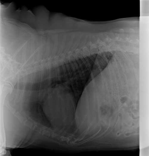
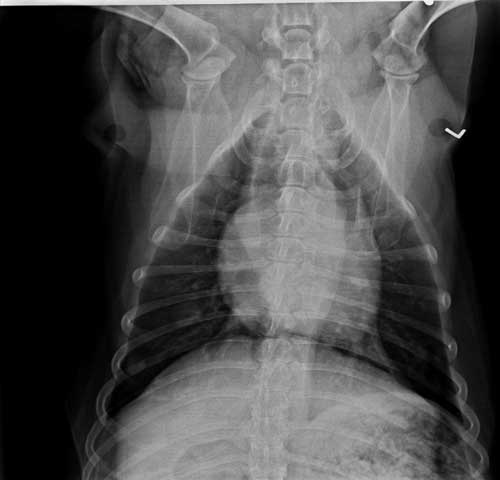
Before any diagnostic or therapeutic steps are taken, the health status of the dog must be fully assessed. Bloodwork and urinalysis should be done to identify any presurgical abnormalities. Thoracic radiographs (both right and left lateral and ventrodorsal planes) should be obtained to search for pulmonary metastases. A fine needle aspiration cytology of the mass is usually not recommended because its diagnostic value to discern between malignant or benign tumors is very low. Regional lymph nodes (lymph glands) should be palpated carefully; fine-needle aspiration or surgical removal are necessary to determine the presence of metastases. Mammary tumors are benign or malignant is to surgically remove them and do a biopsy. It doesn’t matter how many mammary tumors a dog has: because all of them can be different, every mass should be submitted to the lab and analysed. Depending of the size and the number of tumors conservative surgery (lumpectomy/ single mastectomy) or a more aggressive surgery with removal of a whole mammary chain may be recommended.
Prognosis
there is no proven efficacy of any chemotherapeutic protocol for the treatment of malignant mammary tumors in the dog. Certain drugs used for the treatment of carcinomas or sarcomas like gemcitabine/carboplatin/ doxorubicin may be helpful in delaying recurrence or metastases, but their efficacy is unknown.
The prognosis for dogs with malignant mammary tumors depends on the following factors: tumors type, size, regional lymph node (lymph gland) involvement, presence or absence of distant metastases, completeness of resection, local behaviour, vascular or lymphatic invasion, and tumors differentiation.
Histological grading is a good parameter to stratify tumors according to their biological aggressiveness. The Elston and Ellis grading method in humans, invasive ductal breast carcinomas and other invasive tumors are routinely graded. The “Elston and Ellis method” is the most common histological grading method for invasive carcinomas and shows a strong correlation with prognosis.,
Surgery remains the basic treatment for dogs and cats with most type of mammary gland tumors. The exceptions are inoperable disease (e.g., inflammatory carcinoma of the dogs) and distant (organ) metastasis. One of the major problems in veterinary oncology is accurate prognosis for post-surgical mammary cancer cases. In human breast cancer and in canine mammary tumor, histological type, histological grade and lymph node involvement are standard prognostic features. Morphological criteria alone may be insufficient for a proper diagnosis because when only histologically determined, benign tumors may incidentally give rise to metastasis. Despite benign biological behaviour, canine complex adenomas and mixed tumors often show histomorphological evidence of malignancy (carcinoma or sarcoma in benign tumor). Tumor grade and degree of invasion (stage) are also of prognostic significance.
History: – There was a small growth on the ventral abdomen at the level of last pair of teats from the past 2 years, which got enlarged with time. Animal was previously treated at other hospitals too.
|
Clinical Examination |
Specific examination |
Diagnosis | |
| Breed: Labrador. Age: 9 years Body Weight: 29kg
Temperature: 102.9F Heart rate: 125bpm Pulse rate : Respiration rate: Hydration status: Normal CMM: slightly congested |
Blood |
Mammary |
|
| Haematology
Hb:18.5g% PCV:57.5% TLC:12.8*109/L TEC:8.75*1012/L DLC:L%=9.3 M%=4.1 G%=86.6
|
Biochemical
ALT:29.6U/L AST:38.1U/L ALP:275U/L GGT: BG: Bilirubin:1.1mg/dl BUN:40.3mg/dl CRTN:0.4mg/dl TP:
|
Pre medication |
|
| Inj.Butrum 3.2ml i/mInj Atropine 2ml s/cInj Diazepam 3.2ml i/vInj. Intacef Tazo 325 i/vInj. Melonex 1ml i/mInj. Propofol 10ml induction +3ml(top up) Maintenance |
|||
| Clinical/ X-ray and USG
Large pedunculated, fluctuating, warm, firm, multilobulated growth on ventral abdomen at the level of last pair of teats. Small multilobulated growth palpated at level of preceding left teat.
|
Treatment | ||
| Surgical excision of the tumour
9/10/18 Tab Mymox Tab Carodyl Tab Pantroprazole Syp. Hepamust Syp. Haemup 23/10/18 Patient presented for chemotherapy and suture removal. Inj. NSS Inj. Doxorubicin Inj. Vincrystin |
|||
|
The content of the articles are accurate and true to the best of the author’s knowledge. It is not meant to substitute for diagnosis, prognosis, treatment, prescription, or formal and individualized advice from a veterinary medical professional. Animals exhibiting signs and symptoms of distress should be seen by a veterinarian immediately. |




Be the first to comment