Introduction
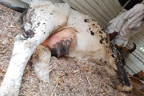
Mastitis is a potentially fatal inflammation of the cow’s mammary gland, which is usually caused by bacteria entering the teat canal and moving into the udder tissue. Toxins released by mastitis bacteria damage milk-secreting tissue and ducts throughout the mammary gland, reducing milk yield and quality. Mastitis can occur at any stage of lactation, including the dry period, but is most likely in the first month after calving and in late lactation.
What Causes Mastitis in Cows
Mastitis occurs when large numbers of white blood cells (leukocytes) migrate into the mammary gland, usually in response to bacteria invading the teat canal through environmental contact or during the milking process.
Streptococcus agalactiae is the most common cause of subclinical mastitis and may rarely cause acute mastitis. The organism lives inside the cow’s udder and survives only for a short time outside the mammary gland. It spreads primarily during milking via the milking machine and contaminated hands and materials (cloths). The microorganism can be eradicated from the herd by appropriate treatment combined with good milking practices
Staphylococcus aureus lives inside and outside the udder on the teat skin. The microorganism can cause both clinical and sub-clinical mastitis and spreads the same way as S. agalactiae.
Yeasts are also responsible for causing mastitis. Overuse of antibiotics and poor sanitation contribute to yeast mastitis. It can also be caused by an injury to the cow’s udder.
Factors Influencing Susceptibility to Mastitis
- Type of bacteria: Influences the type of symptoms and disease. Some bacteria are more virulent than others in causing mastitis.
- Physiological status of cow: Although infection can occurs at any time, most of the new infections take place during the first three weeks of the dry period and during the first month after parturition, suggesting that level of milk production is not directly related to mastitis. It is likely that intra-mammary pressure is a predisposing factor for mastitis during these periods.
- Age of the cow: The incidence of mastitis increases with age. It is possible for the udder of the first-calf heifer to be infected at parturition.
- Level of milk production: Not directly related to incidence of mastitis. However, other factors, which affect milk, yield such as milking rates, pendulous udders may be related to mastitis incidence.
- Inherited features of the cow: Length of the leg in proportion to the udder size and relative strength of the udder attachment are the examples of inherited factors that influence the occurrence of mastitis. Large, pendulous udders tend to exceed the capacity of the supporting ligaments, resulting in breakdown of the udder that will ultimately subject the udder to more physical injuries and thus increases the incidence of mastitis.
- Milking machine: Improper use of milking machine (irregular fluctuation of vacuum level, over-milking, and incomplete milking) is related to tissue irritations and incidence of mastitis.
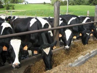
- Environment: Mastitis often increases when cows are turned onto pastures. Housing as it relates to the degree of udder and teat injury are some contributing factors.
Types of Mastitis
There are several ways of classifying mastitis but a simple classification recognizes it into two major groups:
- Contagious Mastitis: Caused by bacteria living on the skin of the teat and inside the udder. It can be transmitted from one cow to another during milking.
- Environmental mastitis: Is caused by the certain organisms such as Escherichia coli which do not normally live on the skin or in the udder but which enter the teat canal. When the cow comes in contact with contaminated environment .
Contagious mastitis can be divided into three groups
- Clinical Mastitis
Characterized by the presence of gross inflammation signs (swelling, heat, redness, pains).
- Peracute Mastitis
Is usually characterized by visibly gross inflammation, disrupted functions (reduction in milk yield, changes in milk composition) and systemic signs (fever, depression, shivering, loss of appetite and loss of weight).
- Acute Mastitis
Similar to peracute mastitis, but with lesser systemic signs (fever and mild depression).
- Sub-Acute Mastitis
In this type of mastitis, the mammary gland inflammation signs are minimal with no visible systemic signs.
- Sub-Clinical Mastitis
Mastitis characterized by change in milk composition with no signs of gross inflammation or milk abnormalities. Changes in milk composition can be detected by special diagnostic tests
- Chronic Mastitis
An inflammatory process that exists for months and may continue from one lactation to another. Chronic mastitis for the most part exists as sub-clinical but may exhibit periodical flare-ups, sub-acute or acute form, which last for a short period of time.
Environmental mastitis
Environmental organisms are found in bedding, soil, walkways, on pasture or any surface with which the cow or her manure comes in contact. Organic bedding materials such as wood shavings, straw or recycled dry manure solids are common sources. Cattle that congregate in cool, shaded areas while on pasture may contaminate the ground with manure and other discharges, thus making this area a source of mastitis causing bacteria.
The prevention of mastitis can be achieved by:
- Proper milking hygiene. Bacteria are transmitted to the udder from the contaminated hands of the milker. Thus the milker’s hands should be washed thoroughly with disinfected soaps before milking while the clinically infected cows should be milked last. Teats should be cleaned and dried before milking.
- Milking machine. Machines for milking should function and operate properly. Vacuum level in the milking unit should be between 300 mm of mercury with little fluctuation. The vacuum regulator should be kept clean and checked regularly.
- Dipping the teats after milking. Teat dipping does not reduce existing infection. However, when suitable disinfectant is used to immerse or spray the teats the rates of new infection can be reduced by up to 50%.
- Dry treatments. Incidence of mastitis during the dry period can be considerably reduced by effective use of antibiotic infusions in each quarter of the udder at the last milking of lactation. Dry cow therapy is the one of the best way to cure chronic and subclinical mastitis that are difficult to treat successfully during lactation.
- Culling of chronically infected cows. This is an effective method because in most herds only 6-8% of all cows account for 40-50% of all clinical mastitis.
- Nutrition. Deficiencies of selenium and vitamin E in the diet have been associated with an increased rate of new mammary infections.
Diagnosis of Mastitis
Current trends in diagnosis of mastitis involves following routine diagnostic tests:
- Physical examination of udder: It is emphasised to view the shape, size, consistency and contour of the udder properly. Detailed examination of the teat and teat orifices should be made to assess inflammation, hot painful swelling and loss of
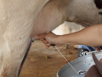
- Strip cup test: In individual animals and in herds, strip cup or strip plate test is routinely used in milking parlor for detection of clinical mastitis. In herd health management practice operators of the milking machines visually examine the fore milk for gross abnormalities by squirting few stripes of milk on strip cup where the abnormalities are usually manifested in the form of blood, flakes, clots and wateriness suggestive of mastitis.
- California mastitis test: It estimates the number of somatic cells present in milk. Procedure for conducting CMT test is by mixing the test reagent (CMT reagent) with an equal quantity of milk. The reagent reacts with DNA of the nuclei of the somatic cells in the milk to form a gel. Depending upon the amount of gel formation. Formation of more gel indicates higher somatic cell count.
- Wisconsin mastitis test: Is primarily a laboratory test which is generally conducted on bulk tank milk samples. In both WMT and CMT same type of reagent is used. WMT the test result reaction are measured (mm). The test is conducted by combining a measured quantity of milk with an equal amount of reagent. The milk and reagent are then mixed for 8 to 10 seconds. The mixture is drained for a period of 18 seconds and returned to an upright position. After waiting for one minute, the amount of fluid remaining in the tube is measured. WMT scores are generally calculated in millimetres (mm) and used to predict the average number of somatic cells present in the milk.
- Modified white side: Test Increased leukocytic count of milk forms the basis of this test. In mastitis, test shows formation of white flakes, while in normal milk; test result shows formation of milky opaque fluid.
- pH Determination test: The normal pH of milk is 6.4 to 6.8 being isotonic with blood plasma. In mastitis occurring during late lactation and dry period, the concentration of lactose and casein in the milk is reduced whereas sodium chloride and sodium bicarbonate pass into the alveoli from plasma to maintain isotonicity. Therefore during such situations milk becomes alkaline with greater amount of chlorides.
- Chloride test: This test detects presence of increased quantity of chlorides in mastitic milk. Normal milk contains about 0.07 per cent of chlorides. In mastitis there is decreased amount of lactose and increased amount of sodium chloride to maintain the normal milk osmotic pressure hence during inflammation there is increase in the chloride content (> 0.14 per cent).
- Electrical conductivity test. The conductivity of milk may be defined as the property of substances in solution which can ionize and therefore can conduct an electrical current. When the concentration of sodium chloride rises in milk, the conductivity rises proportionately. Therefore, measurement of electrical conductivity is used as a simple physical method to diagnose mastitis. Electrical conductivity of milk can be determined by using a hand held (portable) electrical conductivity meter (milk checker or digital mastitis detector). The EC of milk is expressed in the unit of milk seimens/cm (Ms/cm).
- Somatic cell count of milk. In direct microscopic somatic cell counts, milk smear is made on a clean glass slide in the area of 1 cm stained with 1% methylene blue and 60 fields are examined for the count. The average numbers of cells per field are multiplied by the multiplication factor of the microscope and the value so obtained is considered equal to the number of cells/ml of milk sample.
- N-acetyl-β-D-glucosaminidase test (NAGASE) test. This test is based on the measurement of a cell associated enzyme N-acetyl B-D Glucosaminidase in the milk. A highest level of the enzyme indicates high cell count. NAGASE test is a simple, effective and most reliable for diagnosis of sub clinical mastitis. The activity ranges of NAGASE for normal milk (<0.5 x 10 4 cells /ml) and mastitis milk (1.5 x 10⁴ cells/ml) are 0.0053 and 0.034/ mole /min/ml respectively. Among the all-indirect diagnostic tests, NAGASE activation estimation is found to have the highest accuracy.
- Methylene blue reduction test (MBRT). Methylene blue reduction test measures chemical activities, especially the respiratory activities of the bacteria in the milk. The microorganisms in the milk first remove oxygen by respiration and when all oxygen and some other reducible components of the milk has been removed or reduced, there is change in colour of methylene blue to methylene white.
- Milk Antitrypsin Assay (Maum Test): Milk antitrypsin activity is due to leakage of blood Protease inhibitor into milk and represents increased permeability. This was increased to many folds and the trypsin inhibitor test appeared to be superior over CMT in diagnosing sub-clinical mastitis.
| The content of the articles are accurate and true to the best of the author’s knowledge. It is not meant to substitute for diagnosis, prognosis, treatment, prescription, or formal and individualized advice from a veterinary medical professional. Animals exhibiting signs and symptoms of distress should be seen by a veterinarian immediately. |

Be the first to comment