Introduction
Transmissible venereal tumor (TVT) also known as venereal granuloma, infectious sarcoma, Sticker’s sarcoma and canine condyloma is an unusual reticuloendothelial tumor that occur in all dog breeds and is seen in various parts of the world, especially in tropical and subtropical zones. However, it is particularly common in stray dogs, which mate without any control after reaching sexual maturation and is mainly transmitted through coitus [1]. The tumor is primarily located on the genitalia or, less commonly, on the lips or other portions of the skin or mucosa that come in contact with the genitalia. Dogs of both the sexes and all ages are affected, but tumor is most commonly seen in female dogs that have reached sexual maturity. These tumors grow rapidly at first and then remain static for a time, with eventual spontaneous regression after several months. There is infrequent metastasis to regional lymphnodes and rarely to viscera
Transmissible venereal tumors (TVT) are tumors that arise from the dysregulated growth of cells called histiocytes. Histocytes are a type of immune system cell found in many areas of the body, including the skin. TVTs develop from skin histiocytes. TVTs are malignant (cancerous) tumors. Different from other cancers, TVTs are transferrable between dogs. It is sexually transmitted through direct skin-to-skin contact with the tumor that results in cancer cells being transplanted from dog to dog.
Aetiology and transmission
The transmission of TVT by means of intact neoplasm cells was assumed already from the beginning of the 20th century. Later, it was proven that the dog is the only host of TVT. Then it was confirmed that the mutated neoplasm cell itself was the causal agent of TVT and that it induces an immune response in the host. Significant morphological differences have been repeatedly demonstrated to exist between normal dog cells and cells of the tumour. These include constantly some highly specific chromosome aberrations, which, when associated with other factors, lead to the conclusion that TVT derives from cells that have undergone a mutation caused by a still unknown factor. Tumour cells are exfoliated and transplanted during coitus from animal to animal and perpetuate themselves like any other heterologous, mono-cellular organism. The mechanism allowing the neoplastic cells to override nature’s histocompatibility barrier is unknown, while the presence of active immunity has been demonstrated.
Clinical observations
The clinical appearance of TVT has been repeatedly described and is well known. Findings from the majority of cases, recorded in our clinic are summarized below:
Gender and age of infected dogs
Females were infected more often than males (64.5% and 35.5%, respectively). The disease usually (80%) occurred in animals of reproductive age (2 to 8 years old) and less often at older ones.
Main duty- use and social habits of dogs
The main depots of TVT are considered to be populations of unsupervised stray and semi-stray dogs and owned dogs with vague symptoms. Any close contact with those dogs increases the risk of infection. High-risk groups included especially the habitually yard-escaping dogs (75.5%), usually guard dogs (41.3%) and hunting dogs (41.5%), which often came in proximity at the hunters meeting points. TVT was rare in “strictly” supervised home-kept companion animals (4%), occurring only after escape to experience an uncontrolled-unwanted copulation.
Tumour location
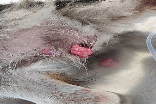
In female dogs the neoplastic lesions were usually located at vestibule and less often at the vagina or invading the vulvar lips. Main lesions were almost always present at the junction of the vestibule and vagina, perhaps due to the high pressure exerted on this area during mating.
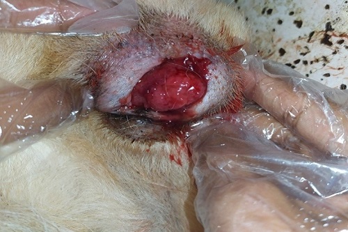
In male dogs neoplastic lesions were usually located on the more caudal part (bulbus glandis) and less often on the shaft (pars longa glandis, or the tip) of the glans penis. Neoplastic lesions, when detected on the preputial mucosa (internal lamina) were always concurrent with those of the glans penis, in contrast to others reports.
Common symptoms
Tumors can appear as small papules or nodules, and over time, they can progress into a cauliflower-like, multi-nodular, or multi-lobulated appearance. They can range in size from 5 mm to 15 cm in diameter, and are usually firm. Tumors often ulcerate, become inflamed, and bleed easily. Other signs associated with these tumors include: The commonest clinical signs observed included a sero-sanguineous or pure hemorrhagic vaginal or preputial discharge
- Bloody discharge from vaginal areas in females
- Bloody discharge from penis in males
- Protrusion of the neoplastic lesions and deformation of the external genitalia The peculiar odour of the neoplastic lesions discharge, which after secondary bacterial infection became particularly unpleasant, and the excessive licking of the genitalia were also signs often reported by keen owners.
Other less common symptoms were dysuria, weakness, ulcers in the perineum area, anorexia constipation, paraphimosis, mating refusal and weight loss. It is clear that clinical findings were less striking in male dogs and the disease was consequently less easily perceptible than in females. Furthermore, in male dogs, there are cases where TVT was accidentally found during other clinical examinations.
Clinical findings as well as haematological and biochemical evaluations strongly suggest that the general health of affected animals was not impaired unless the tumour became necrotic and infected or occluded the urethral orifice, or metastasised.
Hematocrit values were slightly lower than normal in less than 10% of the affected dogs, but no severe anaemia was found, as also reported by other authors. The white cell count was higher than normal in about 30% of the cases; most dogs showed a mild to moderate leukocytosis, probably caused by the inflammation of the tumour surface.
Tumour growth and spread
The time from infection to admission of the dog seems to influence the size of the tumour. During the first six months after infection, the local genital expansion of the neoplasm differed among animals. Thus, there were cases in which the neoplasm was barely perceptible and others in which it expanded and severely corrupted the external genitalia.
The long-existing untreated cases (presenting only mild symptoms) it was found that the extent of neoplastic lesions on the genitalia was closely related to the time elapsed from the onset of the disease (i.e., the neoplasm continued to grow). More specifically, TVT appears to progressively expand until covering most of the external genitalia. Then, it develops more slowly or it may show alternate periods of progressive growth and partial regression. According to our observations on naturally occurring TVTs, none of them had shown spontaneously a complete regression but they persisted at least for a period of 3-4 years without severe consequences to the animal’s health. The diversity in the local distribution or extension of the neoplastic lesions on the genitalia was not found related to factors like animal’s breed, age, gender, usage, or general health condition.
Sometimes the course of the disease was rapid, dramatic or even fatal in metastatic TVT cases. Such severe incidences appeared more frequently in stray, distressed or unhealthy animals, possibly because of a reduced immune response. Common examples refer to animals, which were simultaneously suffering from immunosuppressive diseases (e.g., ehrlichiosis) and/or were subjected to immunosuppressive doses of corticosteroids.
Some dogs that had been definitely cured from TVT by radiotherapy and/or chemotherapy became re-infected two or more years later. This fact leads to the conclusion that precedent TVT infection does not provide long-term immunity.
Treatment
At present, surgical treatment, chemotherapy and radiotherapy are used to treat TVT, while immunotherapy has not been proven effective.
Surgical treatment has been applied since the last century with a low rate of efficacy. The use of electrocautery makes the operation easier and seems to be a little more effective; however it is still far from being suggested as the first choice. Therefore, surgical treatment might be applied to those dogs that present solitary, small, easily accessible and non invasive tumour nodules.
TVT has been proven to be highly sensitive to irradiation, already from the last century. Dosage recommendations range from 1500 to 2500 rads (depending on the chronicity and the extension of neoplastic lesions), divided in sessions of 400-500 rads over a period of 1-2 weeks, or a single dose of 1000 rads which, if not curative, can safely be repeated 1-4 times. However, radiotherapy lacks practicality due to requirements like trained personnel, specialized equipment and expenses. Therefore its use is recommended in cases where other treatments fail.
The intravenous administration of vincristine at the dose of 0.025 mg/m2 of body surface, once a week, for 4-5 weeks, is the treatment of choice, irrespective of a) the neoplasm size–extent; b) the presence of metastases; and c) the duration of the disease. The time needed to complete treatment and the expenses involved are within reasonable limits. The animals fully recover, with no impact on behavior and reproductive efficiency.
Dogs being infected for less than 1 year, i.e., TVT at initial stages of progression are easily treated. The presence of metastasis, the gender or the age of the animals treated do not influence the duration of chemotherapy. Metastasis subsidence concurs impressively.
Chronic cases infected for more than 1 year may resist to treatment, thus demanding therapy of longer duration without ensuring successful results. In case of failure, radiotherapy gives excellent results; alternatively doxorubicin chemotherapy may be applied.
The presence of small size tissue remnants (of less than 0.5 cm), which do not bleed after being rubbed, must not be a reason to continue treatment.
Temporary side effects (partial anorexia, mild depression) may be reported in less than 20% of the treated dogs, usually 1-2 days after vincristine administration. Chemotherapy may cause a decrease of the white blood cell count, but only a few cases present such a leukopenia that might deserve an adjunctive antibiotic treatment or impose suspension (discontinuation) of one or more chemotherapeutic administrations.
Before initiating vincristine chemotherapy, it is important to assess the general health condition of the animal while during therapy it is necessary to follow up the total number of leukocytes, at least at weekly intervals
|
The content of the articles are accurate and true to the best of the author’s knowledge. It is not meant to substitute for diagnosis, prognosis, treatment, prescription, or formal and individualized advice from a veterinary medical professional. Animals exhibiting signs and symptoms of distress should be seen by a veterinarian immediately. |


Be the first to comment