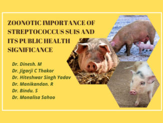
Abstract
Streptococcosis is a group of infectious factorial diseases that affect mainly young animals of many species, caused by pathogenic streptococci and manifested in acute by septicemia and omphalitis, and in subacute and chronic – primary lesions of the lungs, joints, eyes and other organs. Streptococcus dysgalactiae is the second most emerging pathogen causing meningitis, septicaemia, arthritis and sometimes endocarditis in weaned pigs and humans. The primary source of the infection in the newborn piglets is acquired through the infected or carrier post parturient sow. Streptococcus dysgalactiae is diagnosed based on history, clinical sign and gross and hostopathological findings. Further, to confirm the exact cause of disease laboratory technique are mandatory such as gram staining, isolation of bacteria, molecular test (PCR) and biochemical test. Streptococcus dysgalactiae is considered as non-pathogenic for several years, but now perceived as an emerging zoonotic pathogen posing veterinary and public health importance
Introduction
The major hurdle in the growth of pig farming is mainly due to outbreak of various diseases affecting the health and productivity that leads to great economic loss to the farmers. Among the infectious pathogens affecting the pigs, Streptococcosis is an alarming and emerging problem causing considerable damage to swine enterprises (Terekhov et al., 2020). Streptococcosis is a group of diseases that affect mainly young animals of multi-host pathogen due to various species of pathogenic Streptococci. As per recent published report, Streptococcosis is the prime cause of economic losses, particularly in industrial pig production due to high morbidity and mortality. Pathogenic streptococci have both epizootic and epidemiological significance, because of their widespread prevalence with pathogenic potential in both animals and humans. (Laishevtcev et al., 2018; Terekhov et al., 2020).
Without the appropriate treatment, the mortality may reach up to 20% causing a huge economic loss to the farmers. Among the various Streptococcal pathogens, Streptococcus suis, Streptococcus dysgalactiae and Streptococcus porcinus are frequently reported to cause high morbidity and mortality in swine population (Kasuya et al., 2014). In a recent report, S. dysgalactiae (two subspecies), S. porcinus, S. suis and S. pyogenes were the major pathogens associated with diseased conditions in pig breeding farms of Russia (Terekhov et al., 2020).
Etiology
Among the various Streptococcal pathogens, Streptococcus dysgalactiae is considered as emerging pathogen with worldwide increasing significance due to its increased isolation rate from the clinical cases in both human and pigs. Streptococcus dysgalactiae is a Gram positive, pyogenic streptococcal pathogen of humans and animals (Chauhan et al., 2020) It is mainly divided into two subspecies, first is Streptococcus dysgalactiae subspecies dysgalactiae (SDSD) and second is Streptococcus dysgalactiae subspecies equisimilis (SDSE) (Kasuya et al., 2014; Preziuso et al., 2010). SDSD is α, β and non-haemolytic strain of animal origin, belongs to serogroup C and L whereas SDSE is β-haemolytic strain of human and animal origin, presenting mainly group C of and G Lancefield antigens (Vandamme et al., 1996; Kasuya et al., 2014; Leitner et al., 2015; Wajima et al., 2016). Streptococcus dysgalactiae is the second most emerging pathogen causing meningitis, septicaemia, arthritis and sometimes endocarditis in weaned pigs and humans (Oh et al., 2018).
Epidemiology
SDSE is an emerging bacterial pathogen of humans and swine causing wide ranges of diseases (Oh et al., 2018). Considered as non-pathogenic for several years, SDSE has now perceived as an emerging zoonotic pathogen posing veterinary and public health importance (Brandt and Spellerberg 2009). SDSE infection in humans is increasing similar to Streptococcus pyogenes observed over the last decade (Leitner et al., 2015; Babbar et al., 2018). Many environmental factors may affect the incidence of the streptococcosis, such as alterations in feeding management, hygienic parameters, health status of animal, and the load of primary viral or parasitic pathogens (Shabeikin et al., 2018; Terekhov et al. 2020). The predilection site of this pathogen in carrier animals is reported in the palatine tonsils, upper respiratory tract, nasal cavity, digestive tract and genitourinary tract. The primary source of the infection in the newborn piglets is acquired through the infected or carrier post parturient sow. Moreover, the horizontal transmission through contact, aerosol, and alimentary are the contributing factor the widespread prevalence of the pathogen. The sporadic cases of outbreaks have been reported in pigs leading to 50% morbidity and mortality in piglets, which can be easily treated by the administration of beta-lactam antibiotics (Terekhov et al., 2020). Molecular epidemiology plays important role to trace the emergence and possible routes of transmission among humans and animals (Silva et al., 2015).
Economic significance
The swine industry is an integral part of global food security, agricultural economics along with local and international trade across the world (Vanderwaal et al., 2018). As per 20th livestock census, total pig population in India is 9.06 million during Livestock census, 2019 which is decreased by 12.0% as compared to the previous census in 2012 (DAFD, 2019). This decrease in total swine population over the time might be due to infectious diseases primarily constraining swine production as well as emerging and endemic diseases affecting the swine health, which threatens global swine industry with significant economic loss in terms of mortality, production loss, decrease in market value, and restrictions in trade with food insecurity (Vanderwaal et al., 2018).
Pathogenesis
Entry into body of susceptible host (source-skin wounds, the navel or tonsils, post parturient sow)
![]()
Adhesion
![]()
Colonization
![]()
Invasion
![]()
Interact with leukocytes as well as endothelial cells
![]()
Invades to different organs
![]()
Causing severe inflammation and damage to the cells in different organs
![]()
Cell death due to release of free radicles, cytokines
Clinical sign
dysgalactiae subsp. equisimilis causes sporadic infections pigs particularly in recently weaned piglets. It is mainly involved in the cases of septicemia, meningitis, pneumonia, endocarditis and arthritis in growing pigs (Oh et al., 2018, 2020; Pérez-Sancho et al., 2017). These sign of SDSE in pigs are highly overlapping with that of humans (Oh et al., 2020). The clinical sign of SDSE is broadly classified into three forms such as respiratory form, meningitis form and articular form. The meningitis form is mainly observed among the weaned and growers i.e. average of 25-30 days of life. The main clinical signs noted are loss of coordination, convulsive movements, inability to stand, paddling, tremor, and failure to drink and eat food. Many animals become immobile, thus supposed to be dead. The clinical signs exhibited in the meningitis form is as follows: loss of appetite, hyperemic skin, pyrexia, dullness, ataxia, lameness, paralysis, “rowing” movements of the limbs, trembling and convulsive movements. The animals with articular show the pathological process in the joints. Therefore, the movement is restricted their movements, due to which animals are stunted due to less intake of food (Terekhov et al., 2020).
Pathology
Pathology of SDSE infections depends upon the duration of infection and its distribution of infectious pathogens in body. Typical pathological changes were the lesions (gross and microscopic) observed in brain, lungs, heart, joints and spleen suggesting the septicemia. In slaughterhouse survey, S. equisimilis is reported to cause verrucous endocarditis in pigs without any clinical signs of disease. In one earlier report, a female breeding Yorkshire pigs naturally infected with SDSE showed the swollen right tarsal joint with accumulation of cloudy effusion in the articular cavity. The pleura of 6th costovertebral joint showed an abscess of approximately 2 cm in diameter. In another report, 2 piglets showed yellowish discoloration with turbid materials and redness in the femoral synovial cavity along with moderate to severe periarticular fibrous proliferation. Besides joint involvement, enlarged inguinal lymph nodes with serous atrophy of fat were also observed. The lungs infected with Streptococcosis reveal the lesions of fibrinous hemorrhagic pneumonia. However, fibrinous or suppurative bronchopneumonia, interlobular and emphysema, bronchitis, pleurisy are also noticed in the infected piglets (Laishevtcev et al., 2017). The interstitial pneumonia of lung is considered as a significance of the septicaemia caused by SDSE. The infiltration of the meninges by neutrophils, cerebral edema and diffuse purulent meningitis are observed in the pigs naturally affected with SDSE. The microabscess containing abundant polymorphonuclear cells were also noticed in the brain of young piglets affected with SDSE. The purulent-fibrinous pericarditis, hemorrhagic-necrotizing myocarditis and valvular endocarditis are also observed in cardiac involvement. In the articular form, arthritis is observed with hyperemic condition of synovial blood vessels with thickened capsule. The turbid synovial fluid, articular cartilage necrosis (15–30 days post-infection), periarticular fibrosis, abscesses and synovial villi hypertrophy are common necropsy finding in joint form. Moreno et al., (2016) isolated from articular abscess, urine and vaginal discharge of pigs from swineherds of São Paulo State, Brazil from 2007 to 2012.
Humans infected with SDSE infections exhibit the clinical signs of pneumonia, endocarditis, septic arthritis, acute rheumatic fever, acute glomerulonephritis, meningitis and septicaemia (Lo et al., 2015; Oh et al., 2018). Skin and soft-tissue infections are particularly common in cases with S. dysgalactiae subsp. equisimilis bacteremia (Broyles et al., 2009). The frequent finding reported is cellulitis and septic arthritis ( Rantala et al., 2009; Takahashi et al., 2010). Bacteremia in combination with necrotizing fasciitis, pneumonia, and septic shock is life threatening. STSS and necrotizing fasciitis are frequently reported in SDSE bacteremia (Rantala et al., 2014).

Diagnosis
SDSE is diagnosed based on history, clinical sign and gross and hostopathological findings. Further, to confirm the exact cause of disease laboratory technique are mandatory such as gram staining, isolation of bacteria, molecular test (PCR) and biochemical test. Streptococcus genus, are gram-positive bacteria and arranged in chain form which can be seen under microscope after Gram stain. SDSE usually appears on blood agar plates as massive glossy colonies surrounded by a broad zone of β-hemolysis (Ciszewsk et al., 2016; Wajima et al., 2016; Chauhan et al., 2020). The SDSE is catalase and oxidase negative.
Prevention and control
SDSE considered as non-pathogenic for several years, but now perceived as an emerging zoonotic pathogen posing veterinary and public health importance (Brandt and Spellerberg 2009). Many environmental factors may affect the incidence of the streptococcosis, such as alterations in feeding management, hygienic parameters, health status of animal, and the load of primary viral or parasitic pathogens (Shabeikin et al., 2018; Terekhov et al. 2020). Treatment should be started as soon as the disease is diagnosed. Give intramuscular injections of penicillin, cephalosporin or other antibiotics. The strategic use of medication may be applied through drinking water. Use of vaccines may be useful especially in sows before farrowing. Our mothers and grandmothers knew the value of disease prevention after years of respiratory disease and diarrhea in children. Likewise, the sharpest pork producers challenge their veterinarians to provide appropriate disease prevention plans and monitor their results. Disease prevention or control begins with understanding what diseases and risks are present and extends to implementation and monitoring. Careful review of sources and health status lays the groundwork for the future. Following a complete review of health status, control measures are necessary to limit the negative impact of known pathogens. Change must be deliberate and controlled to adequately monitor results. Performance of the growing pig is the ultimate indicator.
Conclusion
SDSE considered as non-pathogenic for several years, but now perceived as an emerging zoonotic pathogen posing veterinary and public health importance. Treatment should be started as soon as the disease is diagnosed. Disease prevention or control begins with understanding what diseases and risks are present and extends to implementation and monitoring. Careful review of sources and health status lays the groundwork for the future.
Reference
- Babbar, A., Itzek, A., Pieper, D.H. and Nitsche-Schmitz, D.P., 2018. Detection of Streptococcus
- pyogenes virulence genes in Streptococcus dysgalactiae subsp. equisimilis from Vellore,
- India. Folia microbiologica, 63(5), pp.581-586.
- Ciszewski, M. and Szewczyk, E.M., 2017. Potential factors enabling human body colonization by animal Streptococcus dysgalactiae subsp. equisimilis strains. Current microbiology, 74(5), p.650.India.
- Department of Animal husbandry, Dairy & Fisheries, M/O Agriculture. Govt of India. 2019. Report. 20th livestock Census.
- Kasuya, K., Yoshida, E., Harada, R., Hasegawa, M., Osaka, H., Kato, M. and Shibahara, T., 2014. Systemic Streptococcus dysgalactiae subspecies equisimilis infection in a Yorkshire pig with severe disseminated suppurative meningoencephalomyelitis. Journal of Veterinary Medical Science, pp.13-0526.
- KATSUMI, M., KATAOKA, Y., TAKAHASHI, T., KIKUCHI, N. and HIRAMUNE, T., 1997. Bacterial isolation from slaughtered pigs associated with endocarditis, especially the isolation of Streptococcus suis. Journal of veterinary medical science, 59(1), pp.75-78.
- KATSUMI, M., KATAOKA, Y., TAKAHASHI, T., KIKUCHI, N. and HIRAMUNE, T., 1998. Biochemical and Serological examination of β-hemolytic Streptococci isolated from slaughtered pigs. Journal of veterinary medical science, 60(1), pp.129-131.
- Kawata, K., Anzai, T., Senna, K., Kikuchi, N., Ezawa, A. and Takahashi, T., 2004. Simple and rapid PCR method for identification of streptococcal species relevant to animal infections based on 23S rDNA sequence. FEMS microbiology letters, 237(1), pp.57-64.
- Kawata, K., Minakami, T., Mori, Y., Katsumi, M., Kataoka, Y., Ezawa, A., Kikuchi, N. and Takahashi, T., 2003. rDNA sequence analyses of Streptococcus dysgalactiae subsp. equisimilis isolates from pigs. International journal of systematic and evolutionary microbiology, 53(6), pp.1941-1946.
- Laishevtcev, A.I., Kapustin, A.V. and Palazyuk, S.V., 2018. Etiological structure of streptococcosis of pigs in various regions of the Russian Federation. Russian Journal of Agricultural and Socio-Economic Sciences, 74(2).
- Laishevtcev, A.I., Kapustin, A.V. and Palazyuk, S.V., 2018. Etiological structure of streptococcosis of pigs in various regions of the Russian Federation. Russian Journal of Agricultural and Socio-Economic Sciences, 74(2).
- Lo, H.H. and Cheng, W.S., 2015. Distribution of virulence factors and association with emm
- polymorphism or isolation site among beta‐hemolytic group G Streptococcus dysgalactiae subspecies equisimilis. Apmis, 123(1), pp.45-52.
- Oh, S.I., Kim, J.W., Jung, J.Y., Chae, M., Lee, Y.R., Kim, J.H., So, B. and Kim, H.Y., 2018. Pathologic and molecular characterization of Streptococcus dysgalactiae subsp. equisimilis infection in neonatal piglets. Journal of veterinary science, 19(2), pp.313-317.
- Oh, S.I., Kim, J.W., Kim, J., So, B., Kim, B. and Kim, H.Y., 2020. Molecular subtyping and antimicrobial susceptibility of Streptococcus dysgalactiae subspecies equisimilis isolates from clinically diseased pigs. Journal of Veterinary Science, 21(4).
- Perez-Sancho, M., Vela, A.I., Garcia-Seco, T., Gonzalez, S., Dominguez, L. and Fernández-Garayzábal, J.F., 2017. Usefulness of MALDI-TOF MS as a diagnostic tool for the identification of Streptococcus species recovered from clinical specimens of pigs. PLoS One, 12(1), p.e0170784.
- Preziuso, S., Laus, F., Tejeda, A.R., Valente, C. and Cuteri, V., 2010. Detection of Streptococcus dysgalactiae subsp. equisimilis in equine nasopharyngeal swabs by PCR. Journal of veterinary science, 11(1), pp.67-72.
- Preziuso, S., Pinho, M.D., Attili, A.R., Melo-Cristino, J., Acke, E., Midwinter, A.C., Cuteri, V.and Ramirez, M., 2014. PCR based differentiation between Streptococcus dysgalactiae subsp. equisimilis strains isolated from humans and horses. Comparative immunology, microbiology and infectious diseases, 37(3), pp.169-172.
- Rantala, S., 2012. A population-based study of beta-hemolytic streptococcal bacteremia: Epidemiological, clinical and molecular characteristics.
- Rantala, S., 2014. Streptococcus dysgalactiae subsp. equisimilis bacteremia: an emerging infection. European journal of clinical microbiology & infectious diseases, 33(8), pp.1303-1310.
- Takahashi, T., Ubukata, K. and Watanabe, H., 2011. Invasive infection caused by Streptococcus dysgalactiae subsp. equisimilis: characteristics of strains and clinical features. Journal of Infection and Chemotherapy, 17(1), pp.1-10.
- Terekhov, P.Y., Matyash, E.A., Yakimova, E.A., Kapustin, A.V., Belyaeva, A.S. and Laishevtsev, A.I., 2020. A new look at the etiological structure of pig streptococcosis. In IOP Conference Series: Earth and Environmental Science (Vol. 421, No. 3, p. 032052). IOP Publishing.
- Vandamme, P., Pot, B., Falsen, E., Kersters, K. and Devriese, L.A., 1997. Taxonomic study of lancefield streptococcal groups C, G, and L (Streptococcus dysgalactiae) and proposal of S. dysgalactiae subsp. equisimilis subsp. nov. International Journal of Systematic and Evolutionary Microbiology, 47(2), pp.605-605.
- VanderWaal, K. and Deen, J., 2018. Global trends in infectious diseases of swine. Proceedings of the National Academy of Sciences, 115(45), pp.11495-11500.
- Wajima, T., Morozumi, M., Hanada, S., Sunaoshi, K., Chiba, N., Iwata, S. and Ubukata, K., 2016. Molecular characterization of invasive Streptococcus dysgalactiae subsp. equisimilis, Japan. Emerging infectious diseases, 22(2), p.247.
- Shabeikin, A.A., Gulyukin, A.M., Belimenko, V.V. and Gulyukin, M.I., 2018. Analysis and assessment of risks of natural focal zoonotic infections outbreaks using geo-information technologies (Moscow; ATT).
- Silva, L.G., Genteluci, G.L., de Mattos, M.C., Glatthardt, T., Figueiredo, A.M.S. and Ferreira-Carvalho, B.T., 2015. Group C Streptococcus dysgalactiae subsp. equisimilis in south-east Brazil: genetic diversity, resistance profile and the first report of human and equine isolates belonging to the same multilocus sequence typing lineage. Journal of medical microbiology, 64(5), pp.551-558.
| The content of the articles are accurate and true to the best of the author’s knowledge. It is not meant to substitute for diagnosis, prognosis, treatment, prescription, or formal and individualized advice from a veterinary medical professional. Animals exhibiting signs and symptoms of distress should be seen by a veterinarian immediately. |


Be the first to comment