Introduction
The Flavobacteriaceae family includes Riemerella anatipestifer. R. anatipestifer is a non-sporulating, gram-negative, catalase and oxidase positive and non-motile. This bacterium was originally classified as belonging to the genera Pasteurella, Pfeifferella or Moraxella, before being reclassified to the genus Riemerella. Currently, at least 21 R. anatipestifer strains that vary in virulence both between and sometimes within a given serotype have been identified. R. anatipestifer infection was first reported in 1932. The infection is a septicaemic (peracute, acute and sometimes chronic) illness that affects geese, ducks, chickens, pheasants, quails, turkeys, guinea fowls and swans. Due to high mortality, the illness causes significant losses in ducks. It affects ducks of all ages, as well as turkeys and chickens, as well as game birds and wild waterfowl. Young ducks have a mortality rate of 2-75 percent. It manifests as acute or chronic septicemia with fibrous polyserositis and meningitis. Because R. anatipestifer has at least 21 recognised serotypes, and distinct serotypes do not give cross-protection against each other, controlling this infection is challenging. More than one serotype is frequently responsible for illness in a flock or on a farm. To give protection against prevalent serotypes, a successful vaccination must be multivalent. Antibiotics can be used to treat the illness, but immunization programmes most effectively manages its outbreak. The infection is not of public health significance and should not be reported to regulatory authorities.
Host/species affected
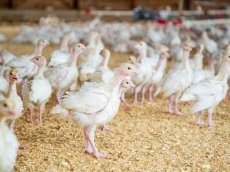
R. anatipestifer infection is mostly a disease of ducks and geese. Turkeys (Helfer and Helmboldt, 1977), chickens (Rosenfeld, 1973), swans (Munday et al., 1970), guinea fowls (Munday et al., 1970) and quails (Zehr and Ostendorf, 1970) can also get affected.
Pathology
Affected birds develop septicaemic lesions as a result of the illness. A yellowish white fluid made up of fibrin coats the heart, liver and air sacs. There are small layers of dry caseous exudate in both the abdominal and thoracic air sacs. The spleen is speckled and the kidneys are swollen. The oviduct includes caseous plugs in females. Lungs are only implicated in a small percentage of cases. Caseous lesions in the nasal and infraorbital sinuses are occasionally seen (Leibovitz, 1972). In ducks, chronic cases of infection is manifested in the joints or the skin. The skin lesions are in the form of dermatitis, with yellowish exudate visible between the fat and skin layers and a spongy, honeycomb-like appearance on the exterior. Turkeys and chickens get comparable lesions, although they are less severe. Fibrinous exudate infiltrated with heterophils and mononuclear cells on air sacs, serous surfaces of the pericardium and liver capsule are histopathological abnormalities. In chronic situations, multinuclear giant cells can be visualized in air sacs. Cloudy edoema, fatty degeneration and necrosis can be presented in the liver cells (Chaudhury and Mohanta, 1985). A rise in the size and quantity of lymph follicles causes spleen mottling. Serofibrinous inflammation of the spinal and cerebral meninges is one of the central nervous system’s lesions.
Epidemiology
Contact through a crack in the skin or with infected birds causes R. anatipestifer infection in the respiratory system. Bacteremia causes systemic infection in the birds. The infection is concentrated in joints and skin in chronic instances. The germs are released into the environment by nasal or infraorbital sinus secretions, infecting feeders and drinkers. In flocks raised in cramped spaces, the virus spreads quickly and kills a lot of young ducklings or goslings. In ducks, there are no seasonal changes in illness incidence. In ducks and geese, outbreaks are prevalent, while turkeys, chickens and other birds have occasional cases. In elderly birds or breeder ducks, the sickness is uncommon. Although mosquitoes have been found to carry R. anatipestifer and may persist in plausible mode, nonetheless no vector has been identified in turkeys (Cooper, 1989).
Clinical symptoms
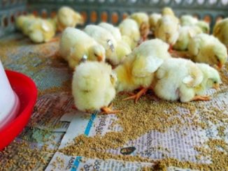
After a 2–5 day incubation period, clinical indications of R. anatipestifer infection generally appear. Ocular and nasal discharges, moderate coughing and sneezing, tremors of the head and neck, weakness, neck tucked-in, ataxia, disinclined to move, incordination, dyspnoea and hyper-excitability are common in affected ducklings, who are normally 1–8 weeks old. In most cases, diseased ducklings lie on their backs, paddling their legs in the latter stages of the disease. Stunting can develop in survivors, with scarring of the air sacs and pericardium leading to slaughter condemnation. The skin and joints are affected by persistent lesions. A yellow-white fluid and congestion can be found throughout the body on post-mortem inspection. Swollen spleen and liver are possible symptoms.
Diagnosis
Clinical examination and postmortem indications can aid in provisional diagnosis. Provisional diagnosis is verified once the organism is isolated and identified – blood or chocolate agar in a candle jar or 5% CO2. Laboratory techniques such as bacterial culture, PCR and ELISA can be used to confirm the diagnosis. R. anatipestifer samples can be treated by inoculating them into 10 ml of Brain Heart Infusion (BHI) broth and incubating them overnight at 37°C in a candle jar under a micro-aerophilic environment.
Molecular confirmation
A PCR test can be used to validate the results at the molecular level. R. anatipestifer species-specific PCR DNA was taken from all of the tentatively identified field isolates and used in a species-specific PCR experiment to determine R anatipestifer. PCR array-based diagnostic tests for R anatipestifer have been reported, and they include assays for the 16S rRNA, rpoB gene, ompA gene and an ERIC fragment. Because of the significant odds of false-positive results, PCR amplification of a small segment of the rpoB or 16S rRNA gene followed by sequencing is recommended to confirm identification.
Differential diagnosis from
- P. multocida, E. coli, streptococcal infections.
- Ducklings infected with salmonella may exhibit anxious symptoms similar to those seen in R. anatipestifer infection.
- Duck chlamydiosis.
Treatment
Sulphonamides (sulfamethazine and sulfaquinoxaline), as well as potentiated sulphonamides, are the medications of choice for Riemerella anatipestifer therapy. It’s included in the water supply. Penicillin + dihydrostreptomycin or streptomycin + dihydrostreptomycin subcutaneous injections are also quite successful. The use of Lincomycin can help to minimise mortality. Lincospectin (a lincomycin-spectinomycin combination) is also indicated for therapy.
Zoonotic potential
Infection with R. anatipestifer has no public health implications. Bird corpses with infection sores at the time of slaughter are often rejected or degraded.
Prevention and Control
In order to obviate R. anatipestifer infection in ducks, standard management techniques are essential. Strict biosecurity must be adhered to. Cleaning and disinfection between flocks, as well as flock separation on multi-age farms, are important considerations. Allowing facilities to dry out and be exposed to hot dry air can help some facilities lower pathogen loads. In the farm, good cleanliness standards should be followed. Stock overcrowding should be avoided at all costs. Cleaning and disinfection to be followed regularly on and around the farm. When possible, employing all-in/all-out management systems, and providing time for downtime between flocks. On most commercial duck and goose farms, bacterin should be given to naïve ducklings and breeder birds. Because of the rise of antimicrobial resistance, commercial operations require immunization and effective biosecurity.
Vaccination
Ducks and naïve ducklings can both be given a live vaccination that includes the three most frequent serotypes of R. anatipestifer (i.e. serotypes 1, 2, and 5). Multiple vaccine manufacturers offer autogenous bacterins for different serotypes. In turkeys, an autogenous oil-emulsion bacterin might be employed. Breeder ducks can be vaccinated with a bacterin or a live vaccine to provide their ducklings protection that lasts up to 2-3 weeks.In certain places, inactivated and attenuated vaccinations are available. Autogenous bacterins are occasionally employed.
Ducklings can be immunized against R. anatipestifer infection using inactivated vaccines (Harry and Deb, 1979; Sandhu, 2003). To give protection, autogenous inactivated vaccinations are routinely employed. Because many serotypes might be found in a single hatch of ducks on a farm or in a given location, vaccinations are frequently multivalent to provide broad-spectrum protection against the disease’s primary serotypes. Bacterial cells are cultured in broth medium and destroyed with formalin to make inactivated vaccines. Some vaccinations are oil-emulsified or contain adjuvants eg. aluminium hydroxide. Due to the fact that most ducks are slaughtered at the age of 6-7 weeks, adjuvanted vaccinations may cause lesions at the injection site, resulting in the condemnation of a portion of the carcass. At 2 and 3 weeks of age, inactivated vaccinations are commonly given subcutaneously in the neck.
When given to one-day-old ducklings, a live avirulent vaccine produced against R. anatipestifer serotypes 1, 2 and 5 infection provides considerable protection (Sandhu, 1991). In the lab, protection was seen up to 7 weeks of age, while in the field, some commercial duck farms use an inactivated vaccination from 2-4 weeks of age. Maternal immunity protects the offspring of breeder ducks that have been vaccinated with an inactivated or live vaccine for up to 2-3 weeks.
References
Available on request
| The content of the articles are accurate and true to the best of the author’s knowledge. It is not meant to substitute for diagnosis, prognosis, treatment, prescription, or formal and individualized advice from a veterinary medical professional. Animals exhibiting signs and symptoms of distress should be seen by a veterinarian immediately. |


Be the first to comment