Introduction
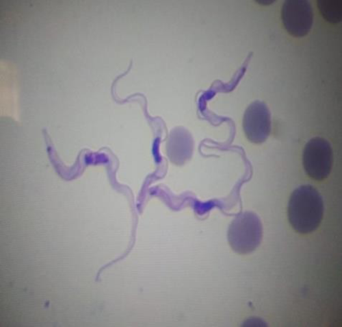
Surra, or animal trypanosomosis, is a tropical and subtropical disease caused by Trypanosoma evansi (a eukaryotic, unicellular, euryxenous, haemoflagellate organism). The disease is endemic throughout India, particularly in low-lying areas. However, disease incidence may be higher during the rainy and post-rainy seasons due to a higher prevalence of tabanid flies, as this period provides the optimal conditions for fly vector propagation. Due to the fact that the disease occurs chronically in cattle and buffaloes as an asymptomatic infection, these carriers may act as a nidus for infection to transmit to more susceptible hosts such as camels and horses, resulting in a high fatality rate. Disease outbreaks in ruminants and other animals have been observed on a routine basis throughout India.
The syndromes associated with this parasite infection are serious, and numerous medicines have been commonly used to treat the infection produced by T. evansi, with drug resistance developing. Genes that influence host range, virulence, host immunity to the parasite, and transmissibility are critical in terms of human and animal disease, highlighting the importance of realizing the pathogenic mechanisms and metabolism of T. evansi infections, particularly in the context of emerging evidence of T. evansi infections in humans. We present a cumulative view of the pathogenic pathways behind T. evansi infection, which are known to promote attrition and so influence host immunity.
Clinciopathology of evansi
The first sign of infection with any Trypanosome strain is a skin swelling (chancre) in which the parasites thrive. Skin edoema is followed by fever, which is directly related to parasitemia. Throughout the course of the disease, recurrent episodes of fever and parasitemia occur, as well as a loss of condition and lassitude. Edema, particularly in the lower portions of the body, might occur late in the course of the infection. Additionally, urticarial plagues and petechial serous membrane haemorrhages are frequently detected. Skin rashes on the ears and lateral side of the body are visible, and abortions have been documented in the majority of susceptible species. T. evansi has been found to infiltrate and spread throughout the central nervous system (CNS), resulting in severe and potentially deadly clinical symptoms during the disease’s second stage. This situation results in an illness that can range from chronic to acute and fatal, manifesting clinically as progressive weakness, emaciation, depletion, recurring fever, enlarged lymph nodes, and death. Variant surface glycoprotein (VSG) coats the Trypanosome’s surface and serves as the primary antigenic determinant for the human immune system. The variability stems from the parasite’s capacity to shed its coat and regenerate it by producing another surface glycoprotein. Due to the successive expression of VSGs, an ever-changing antigenic pattern is produced. Thus, antigenic diversity in T. evansi Trypanosomes appears to be the principal method by which the Trypanosomes evade the host’s immune response. The VSG coat comprises around 10% of total protein, and the genome has 1000 genes capable of expressing VSG genes which are switched on and off randomly during each generation.
The most common clinical manifestation of T. evansi is progressive anaemia caused mostly by haemolysis of red blood cells and erythrophagocytosis. Apart from progressive anaemia and intravascular coagulation caused by the parasite’s toxins and proteolytic enzymes, hypoglycemia is believed to be the cause of mortality. Other findings include a disintegration of the blood-brain barrier and an increase in intracerebral immunoglobulin production. T. evansi-induced immunosuppression leads in the failure of immunisation programmes against bacterial and viral disease and secondary/concurrent infection with additional pathogens . Oxidative damage could be caused by alterations in the activity of antioxidants such as reduced glutathione (GSH), superoxide dismutase (SOD), and catalase (CAT). Additionally, lipid peroxidation has been linked to erythrocyte oxidative damage and anaemia. Additionally, several publications have demonstrated the critical involvement of free radical-induced oxidative stress in the development of trypanosomosis. Trypanosome infection results in the creation of high numbers of reactive oxygen species (ROS) and free radicals (FR), which function as cytotoxic agents, causing damage to important cell components such as proteins and lipids.
Anemia
Anemia is a condition in which the quantity of red blood cells, their volume, or their haemoglobin content are less than normal. Anemia is a frequent finding in patients with clinical trypanosomiasis and trypanosomosis . It appears to be mainly hemolytic in nature, as evidenced by decreased erythrocyte life span and severe erythrophagocytosis Moreover, anaemia has been linked to the development of the majority of tissue degenerative alterations and, consequently, the diseases observed in trypanosome-infected animals In severe infections, the beginning of anaemia is correlated with the presence of trypanosomes in the blood, and the severity is related to the parasitemia level and initial wave. Numerous reports have highlighted hemolytic factors such as hemolysis and free fatty acids, immunologic mechanisms, hemodilution, coagulation disorders, erythrogenesis inhibition, and release of trypanosomal sialidase in the development of anaemia in trypanosomiasis. Anemia is primarily caused by hemolytic crises in both human and animal trypanosomes, in which the erythrocytes are destroyed by an augmented mononuclear phagocytic system.Anemia is generally triggered by the action of sialidase, which cleaves the sialic acids on the surface of erythrocytes, revealing Galactosyl residues. These residues are then recognised by macrophage-specific D-galactose lectins, resulting in erythrophagocytosis and ultimately anaemia.
Conclusion
Anemia persists as the cardinal sign during T. evansi infection, but its cause and kinetics appear to vary depending on the presence or absence of Trypanosomes. T. evansi‘s pathophysiology is complex, and the cause of death is still unknown. Trypanosomes infect their hosts by rapidly replicating in the blood and eventually in the central nervous system (CNS). This requires energy, which the host provides for the Trypanosome’s existence. The Trypanosome secretes phospholipases that hydrolyze the host RBC membrane, as well as sialidases that degrade sialic acid. These events occur as a result of hemolytic programmes that shorten the life of erythrocytes, resulting in anaemia.


Be the first to comment